. 2021 May 25;52(1):73.
doi: 10.1186/s13567-021-00938-6.

SYNCRIP facilitates porcine parvovirus viral DNA replication through the alternative splicing of NS1 mRNA to promote NS2 mRNA formation

Affiliations

SYNCRIP facilitates porcine parvovirus viral DNA replication through the alternative splicing of NS1 mRNA to promote NS2 mRNA formation

Songbiao Chen et al. Vet Res. .

Abstract

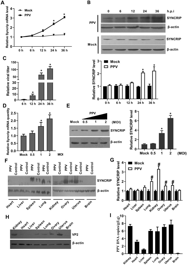
Porcine Parvovirus (PPV), a pathogen causing porcine reproductive disorders, encodes two capsid proteins (VP1 and VP2) and three nonstructural proteins (NS1, NS2 and SAT) in infected cells. The PPV NS2 mRNA is from NS1 mRNA after alternative splicing, yet the corresponding mechanism is unclear. In this study, we identified a PPV NS1 mRNA binding protein SYNCRIP, which belongs to the hnRNP family and has been identified to be involved in host pre-mRNA splicing by RNA-pulldown and mass spectrometry approaches. SYNCRIP was found to be significantly up-regulated by PPV infection in vivo and in vitro. We confirmed that it directly interacts with PPV NS1 mRNA and is co-localized at the cytoplasm in PPV-infected cells. Overexpression of SYNCRIP significantly reduced the NS1 mRNA and protein levels, whereas deletion of SYNCRIP significantly reduced NS2 mRNA and protein levels and the ratio of NS2 to NS1, and further impaired replication of the PPV. Furthermore, we found that SYNCRIP was able to bind the 3'-terminal site of NS1 mRNA to promote the cleavage of NS1 mRNA into NS2 mRNA. Taken together, the results presented here demonstrate that SYNCRIP is a critical molecule in the alternative splicing process of PPV mRNA, while revealing a novel function for this protein and providing a potential target of antiviral intervention for the control of porcine parvovirus disease.

Keywords: Alternative splicing; NS1 mRNA; NS2 mRNA; PPV; SYNCRIP.


Conflict of interest statement

The authors declare that they have no competing interests.

Figures

Figure 1
Figure 1
PPV transcription map. A PPV genome. Linear single-stranded negative PPV genome is shown. ITR, inverted terminal repeat. B Different open reading frames are indicated with different colors. C Different RNA encoding different viral proteins.
Figure 2
Figure 2
Screening and identification of NS1 mRNA interacting host proteins. A Experimental design for pulldown assays and identification of NS1 mRNA associated cellular proteins. NS1 mRNA and LacZ mRNA were biotinylated by transcription in vitro, and incubated with PK-15 total cell lysates. B Coomassie bright blue staining of biotinylated NS1 mRNA associated proteins. C The gene ontology (GO) analysis on NS1 mRNA interacting with host proteins. The GO analysis was performed by mRNA processing of the biological process on NS1 mRNA interacting with host proteins using webgestalt. D HEK293 cells were transfected with different expression vectors containing Flag tagged host proteins for 24 h. RNA-pulldown was performed with streptavidin beads, followed by western blotting using the anti-Flag antibodies. E Western blotting identified the interaction between NS1 mRNA and endogenous SYNCRIP protein in PPV-infected PK-15 cells by RNA-pulldown. F RNA-immunoprecipitation identified the interaction between NS1 mRNA and endogenous SYNCRIP protein in PPV-infected PK-15 cells. An anti-SYNCRIP antibody or negative control IgG was used to pull down RNA–protein complexes. Recovered cDNA from PPV-infected PK-15 cells was examined for viral RNA by PCR with primer sets of NS1 and GAPDH, Y-PPV plasmid was used as a template for positive controls of PCR. G Laser confocal identify the interaction between NS1 mRNA and endogenous SYNCRIP protein in PPV-infected PK-15 cells. H Identification of GST and GST-SYNCRIP protein purification. (I) EMSA identified the interaction between NS1 mRNA and endogenous SYNCRIP protein. Scale bar = 10 μm.
Figure 3
Figure 3
PPV infection up-regulates SYNCRIP expression in vitro and in vivo. PK-15 cells were infected with 1 MOI of PPV or mock infection for the time indicated, the mRNA level of SYNCRIP (A), the protein level of SYNCRIP (B) and the relative viral titers of different infection times were measured by TCID50 (C). D-E qPCR and western blotting analysis of SYNCRIP mRNA and protein levels in PK-15 cells infected with different doses of PPV (MOI = 0, 0.5, 1, or 2) for 24 h. β-actin served as an internal control. F–G Western blotting and qPCR analysis of SYNCRIP protein and mRNA levels in different tissues of PPV infected pregnant sows. H–I Western blotting and q-PCR analysis of viral VP2 protein levels and DNA copies in different tissues of PPV infected pregnant sows. β-actin served as an internal control. The results are shown as the mean ± SD (n = 3). *p < 0.05, versus mock infected cells at same time points. #p < 0.05, versus mock tissues at same tissue.
Figure 4
Figure 4
Knockout of SYNCRIP inhibits the viral replication in PPV‑infected cells. A Schematic chromatogram representation of sgRNA targeting at the SYNCRIP genomic region. PAM sequences are underlined and highlighted in green. sgRNA targeting sites are underlined and highlighted in red. Red arrows indicate gRNA targeting sites. B Western blotting analysis of the SYNCRIP expression in PK-15 cells infected with CRISPR/Cas9 lentivirus and then selected by puromycin. The lentiviruses contain gRNA-63 and gRNA-210. C Cell viability of cell lines stably knockout for SYNCRIP. D-E Knockout of SYNCRIP inhibits PPV progeny virion production. PK-15, PKSYNCRIP+/+ and PKSYNCRIP−/− cells were infected with PPV (MOI = 1) for 24 h. The relative fold-change PPV DNA copies and viral titers were determined by q-PCR and TCID50 assay. The results are shown as the mean ± SD (n = 3). *p < 0.05 versus PK-15 cells with the same treatment.
Figure 5
Figure 5
Over-expression of SYNCRIP leads to the reduction of NS1 mRNA and protein expression. A Western blotting analysis of SYNCRIP protein levels in PK-15 cells with SYNCRIP over-expression using anti-His monoclonal antibodies. PK-15 as the cells without transfection, PKV as the cells transfected with blank vector, PKSYNCRIP as the cells transfected with the vector expressing His-SYNCRIP. B Western blotting analysis of SYNCRIP protein levels in PK-15 cells with SYNCRIP over-expression using SYNCRIP monoclonal antibodies. C Cell viability of cell lines stably overexpressing SYNCRIP. D Western blotting analysis of viral NS1, NS2, VP1 and VP2 proteins expression levels in PK-15 cells with SYNCRIP over-expression. E Western blotting analysis of NS1 protein expression levels in PK-15 cells of SYNCRIP over-expression. F Western blotting analysis of NS1 protein levels in PK-15 cells co-transfected with different doses of SYNCRIP (0, 1, 2, 4 or 8 μg) for 24 h.G Western blotting analysis of NS1 protein levels in ST cells co-transfected with different doses of SYNCRIP (0, 1, 2, 4 or 8 μg) for 24 h. H Q-PCR analysis of NS1 mRNA levels in PK-15 cells co-transfected with different doses of SYNCRIP (0, 1, 2, 4 or 8 μg) for 24 h. I Q-PCR analysis of NS1 mRNA levels in ST cells co-transfected with different doses of SYNCRIP (0, 1, 2, 4 or 8 μg) for 24 h. The results are shown as the mean ± SD (n = 3). *p < 0.05 versus untreated cells with the vector pCDNA-His-SYNCRIP over-expression.
Figure 6
Figure 6
SYNCRIP regulates the ratio of NS1 mRNA to NS2 mRNA and the effect on viral replication. A Laser confocal identification of the effect of SYNCRIP on viral entry in the presence of the translation inhibitor cycloheximide. B NS1 mRNA and its alternatively spliced products are depicted and the arrowheads show primer positions for detection of various mRNA in the following experiments. Cells were infected with PPV, and total RNA was isolated and analyzed by q-PCR with primers specific to NS1, NS2, NS3, VP1 and VP2 mRNA. Ratios of NS2 mRNA to NS1 (C) or to NS3 (D), or VP2 to VP1 (E) at the indicated times are presented. F Diagram of NS2 knockout Y-PPVNS2−. The NS2 protein knockout Y-PPV clone (Y-PPVNS2−) is diagrammed and shown with mutations of three translation initiation codons from CTC to TAG. G Transmission electron microscopy of rescued Y-PPVNS2− virus. Viral particle morphology was observed using transmission electron microscopy. H Q-PCR analysis of parental PPV and Y-PPVNS2− relative DNA copies at different infection time points. I SYNCRIP knockout decreases viral DNA replication via reduction of NS2 protein expression. PK-15 cells were infected with Y-PPV or Y-PPVNS2−, and NS2 proteins were in part of these cells as indicated. PK-15SYNCRIP−/− cells or NS2 protein expressing PK-15SYNCRIP−/− cells were infected with Y-PPV. At 24 h post-infection, the supernatants from each infected sample was collected and used for virion quantification by TCID50. The results are shown as the mean ± SD (n = 3). ns: no significant difference; #p < 0.05 versus PK-15 cells infected with PPV at the same time points. **p < 0.01 versus PK-15 cells with the same treatment.
Figure 7
Figure 7
SYNCRIP affects NS1 expression by acting on the 3′-terminal site of NS2 mRNA. A A schematic diagram of NS1 mRNA mutation site. B Western blotting analysis of the expression of different NS1 mutants in PK-15SYNCRIP−/− cells. C Western blotting analysis of viral parental NS1, NS1pNmut, NS1pCmut and NS1pNpCmut protein expression levels in PK-15 cells with SYNCRIP over-expression. D Western blotting analysis of NS1pCmut protein levels (upper) and mRNA levels (lower) in PK-15 cells co-transfected with different doses of SYNCRIP for 24 h. E Western blotting analysis of NS1pNpCmut protein levels (upper) and mRNA levels (lower) in PK-15 cells co-transfected with different doses of SYNCRIP for 24 h. F Western blotting analysis of NS1pNmut protein levels (upper) and mRNA levels (lower) in PK-15 cells co-transfected with different doses of SYNCRIP for 24 h. G The effects of NS1pCmut mutation on PPV DNA copy number were determined by q-PCR assay. (H) The effects of NS1pCmut mutation on PPV viral titer were determined by TCID50 assay. The results are shown as the mean ± SD (n = 3). **p < 0.01 versus PK-15 cells with the same treatment at the same time points.

References

    1. Johnson RH, Collings DF. Experimental infection of piglets and pregnant gilts with a parvovirus. Vet Rec. 1969;85:446–447. doi: 10.1136/vr.85.16.446. - DOI - PubMed
    1. Meszaros I, Toth R, Olasz F, Tijssen P, Zadori Z. The SAT protein of porcine parvovirus accelerates viral spreading through induction of irreversible endoplasmic reticulum stress. J Virol. 2017;91:e00627–e717. doi: 10.1128/JVI.00627-17. - DOI - PMC - PubMed
    1. Majumder K, Boftsi M, Whittle FB, Wang J, Fuller MS, Joshi T, Pintel DJ. The NS1 protein of the parvovirus MVM Aids in the localization of the viral genome to cellular sites of DNA damage. PLoS Pathog. 2020;16:e1009002. doi: 10.1371/journal.ppat.1009002. - DOI - PMC - PubMed
    1. Niskanen EA, Kalliolinna O, Ihalainen TO, Hakkinen M, Vihinen-Ranta M. Mutations in DNA binding and transactivation domains affect the dynamics of parvovirus NS1 protein. J Virol. 2013;87:11762–11774. doi: 10.1128/JVI.01678-13. - DOI - PMC - PubMed
    1. Lin P, Cheng Y, Song S, Qiu J, Yi L, Cao Z, Li J, Cheng S, Wang J. Viral nonstructural protein 1 induces mitochondrion-mediated apoptosis in mink enteritis virus infection. J Virol. 2019;93:e01249–e1319. doi: 10.1128/JVI.01249-19. - DOI - PMC - PubMed

MeSH terms