

Unilateral acute posterior multifocal placoid pigment epitheliopathy in a convalescent COVID-19 patient
- PMID: 34034832
- PMCID: PMC8148402
- DOI: 10.1186/s40942-021-00312-w
Unilateral acute posterior multifocal placoid pigment epitheliopathy in a convalescent COVID-19 patient
Abstract
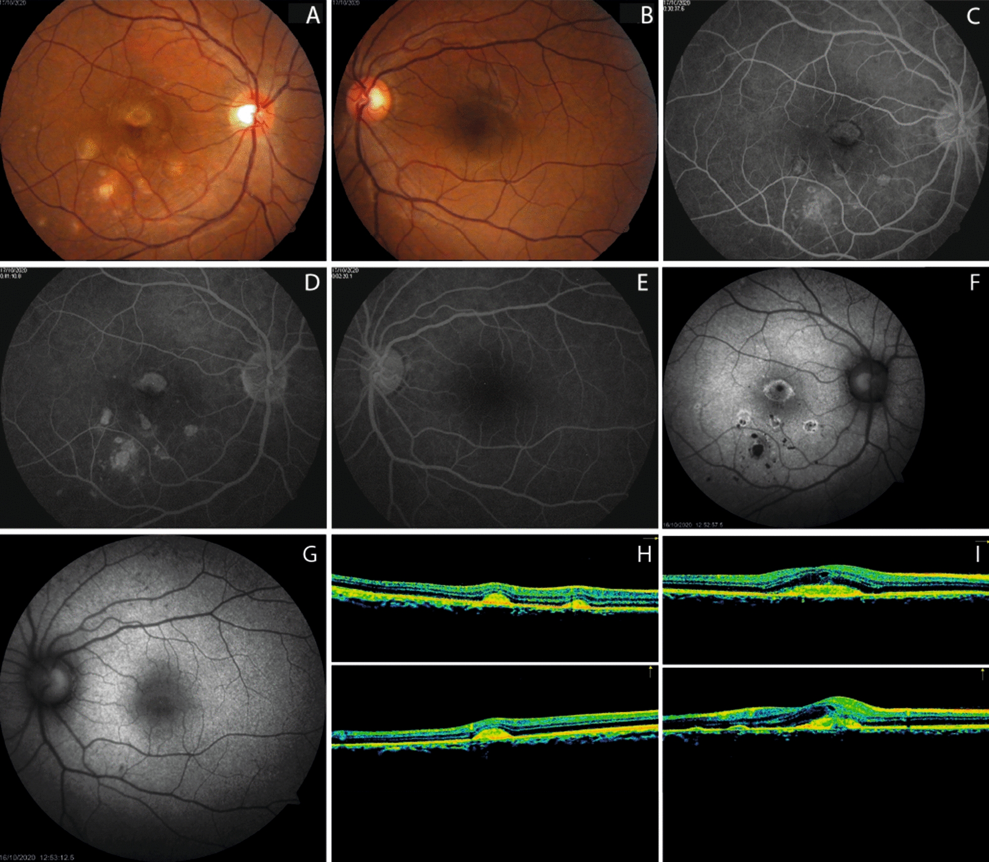
Background: To report a case of unilateral acute posterior multifocal placoid pigment epitheliopathy (APMPPE) in a Hispanic convalescent COVID-19 female patient. Case presentation A 35-year-old Hispanic female with exposure to the severe acute respiratory syndrome coronavirus 2 (SARS-CoV-2) was evaluated due to unilateral visual loss. Ophthalmic examination and diagnostic tests were consistent with APMPPE.
Discussion: Ocular changes can be observed in patients with COVID-19. A complete ophthalmic evaluation must be performed in patients with low vision after SARS-CoV-2 infection.
Keywords: Acute posterior multifocal placoid pigment epitheliopathy; Coronavirus disease 2019; Severe acute respiratory syndrome coronavirus 2.
Conflict of interest statement
The authors declare that they have no competing interests.
Figures
Similar articles
-
Unilateral acute posterior multifocal placoid pigment epitheliopathy (APMPPE) with delayed contralateral eye involvement.BMC Ophthalmol. 2024 Jan 9;24(1):17. doi: 10.1186/s12886-023-03221-8. BMC Ophthalmol. 2024. PMID: 38195467 Free PMC article.
-
Long-term follow-up of a bilateral acute posterior multifocal placoid pigment epitheliopathy following COVID-19 infection: a case report.J Ophthalmic Inflamm Infect. 2024 Jan 4;14(1):2. doi: 10.1186/s12348-023-00382-x. J Ophthalmic Inflamm Infect. 2024. PMID: 38177891 Free PMC article.
-
Acute posterior multifocal placoid pigment epitheliopathy following COVID-19 infection.Am J Ophthalmol Case Rep. 2023 Mar;29:101790. doi: 10.1016/j.ajoc.2022.101790. Epub 2022 Dec 29. Am J Ophthalmol Case Rep. 2023. PMID: 36597447 Free PMC article.
-
Acute posterior multifocal placoid pigment epitheliopathy (APMPPE).J Ophthalmic Inflamm Infect. 2021 Nov 1;11(1):31. doi: 10.1186/s12348-021-00263-1. J Ophthalmic Inflamm Infect. 2021. PMID: 34524577 Free PMC article. Review.
-
[Acute posterior multifocal placoid pigment epitheliopathy. A rare cause of ischaemic stroke].Rev Neurol. 2013 Jun 1;56(11):567-72. Rev Neurol. 2013. PMID: 23703059 Review. Spanish.
Cited by
-
Use of Imaging Technology to Assess the Effect of COVID-19 on Retinal Tissues: A Systematic Review.Ophthalmol Ther. 2022 Jun;11(3):1017-1030. doi: 10.1007/s40123-022-00509-8. Epub 2022 Apr 29. Ophthalmol Ther. 2022. PMID: 35488102 Free PMC article. Review.
-
Atypical Acute Ischemic Choriocapillaritis: A Case Report.Clin Case Rep. 2024 Dec 16;12(12):e9646. doi: 10.1002/ccr3.9646. eCollection 2024 Dec. Clin Case Rep. 2024. PMID: 39691487 Free PMC article.
-
Relentless placoid chorioretinitis: A review of four cases in pediatric and young adult patients with a discussion of therapeutic strategies.Front Pediatr. 2023 Mar 27;11:885230. doi: 10.3389/fped.2023.885230. eCollection 2023. Front Pediatr. 2023. PMID: 37051435 Free PMC article. Review.
-
Immune Privilege Furnishes a Niche for Latent Infection.Front Ophthalmol (Lausanne). 2022 Mar 8;2:869046. doi: 10.3389/fopht.2022.869046. eCollection 2022. Front Ophthalmol (Lausanne). 2022. PMID: 38983514 Free PMC article. Review.
-
Unilateral acute posterior multifocal placoid pigment epitheliopathy (APMPPE) with delayed contralateral eye involvement.BMC Ophthalmol. 2024 Jan 9;24(1):17. doi: 10.1186/s12886-023-03221-8. BMC Ophthalmol. 2024. PMID: 38195467 Free PMC article.
References
-
- World Health Organization, Naming the Coronavirus Disease (COVID-19) and the Virus That Causes It, 2020. https://www.who.int/emergencies/diseases/novel-coronavirus-2019/technica.... Accessed 5 May 2021.
LinkOut - more resources
Full Text Sources
Other Literature Sources
Miscellaneous
