. 2021 May 25;6(59):eabj1750.
doi: 10.1126/sciimmunol.abj1750.

SARS-CoV-2 variants of concern partially escape humoral but not T-cell responses in COVID-19 convalescent donors and vaccinees

Affiliations

SARS-CoV-2 variants of concern partially escape humoral but not T-cell responses in COVID-19 convalescent donors and vaccinees

Daryl Geers et al. Sci Immunol. .

Abstract

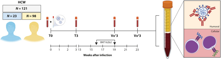
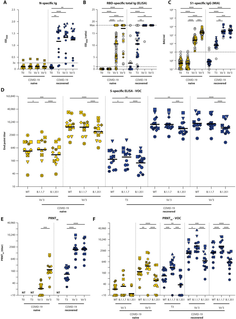
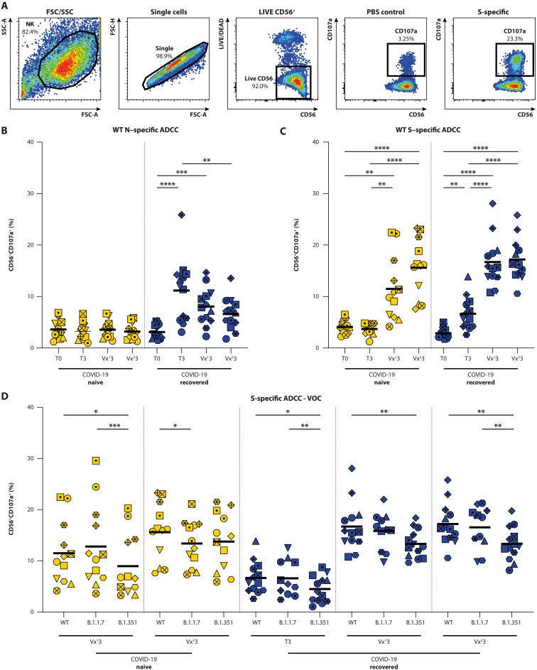
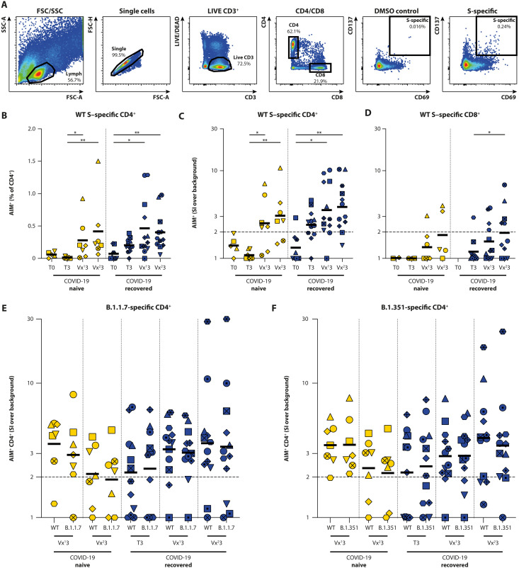
The emergence of SARS-CoV-2 variants harboring mutations in the spike (S) protein has raised concern about potential immune escape. Here, we studied humoral and cellular immune responses to wild type SARS-CoV-2 and the B.1.1.7 and B.1.351 variants of concern in a cohort of 121 BNT162b2 mRNA-vaccinated health care workers (HCW). Twenty-three HCW recovered from mild COVID-19 disease and exhibited a recall response with high levels of SARS-CoV-2-specific functional antibodies and virus-specific T cells after a single vaccination. Specific immune responses were also detected in seronegative HCW after one vaccination, but a second dose was required to reach high levels of functional antibodies and cellular immune responses in all individuals. Vaccination-induced antibodies cross-neutralized the variants B.1.1.7 and B.1.351, but the neutralizing capacity and Fc-mediated functionality against B.1.351 was consistently 2- to 4-fold lower than to the homologous virus. In addition, peripheral blood mononuclear cells were stimulated with peptide pools spanning the mutated S regions of B.1.1.7 and B.1.351 to detect cross-reactivity of SARS-CoV-2-specific T cells with variants. Importantly, we observed no differences in CD4+ T-cell activation in response to variant antigens, indicating that the B.1.1.7 and B.1.351 S proteins do not escape T-cell-mediated immunity elicited by the wild type S protein. In conclusion, this study shows that some variants can partially escape humoral immunity induced by SARS-CoV-2 infection or BNT162b2 vaccination, but S-specific CD4+ T-cell activation is not affected by the mutations in the B.1.1.7 and B.1.351 variants.


Figures

Fig. 1.
Fig. 1.. HCW study design.
N = 121 HCWs were enrolled in a prospective SARS-CoV-2 infection and vaccination study. Upon symptomatic presentation to occupational health services, a paired nasopharyngeal swab and EDTA blood sample was obtained (T0). A second EDTA blood sample was obtained 3 weeks after diagnostic RT-PCR (T3). On the basis of the diagnostic RT-PCR result at T0 and serology result at T3, 98 COVID-19–naive (yellow) and 23 COVID-19–recovered (blue) HCWs were enrolled in the vaccination study on average 50 days after inclusion. N = 13 COVID-19–recovered and N = 12 COVID-19–naive participants were randomly selected for in-depth analysis. Blood samples were collected after the first (Vx13) and second (Vx23) vaccination, processed, and subsequently used for downstream serological and cellular assays.
Fig. 2.
Fig. 2.. Detection of SARS-CoV-2–specific humoral responses.
Total Ig levels were measured in COVID-19–naive (yellow) and –recovered (blue) donors at the acute, convalescent, post-vaccination 1, and post-vaccination 2 stage (T0, T3, Vx13, and Vx23) by an (A) ELISA against nucleocapsid (N) and (B) RBD. (C) Quantitative IgG against S1 was measured by a Luminex bead assay. (D) Antibody binding to WT SARS-CoV-2 and VOC B.1.1.7 and B.1.351 was determined by end point titration in ELISA. Virus neutralization was measured by PRNT50 against (E) WT SARS-CoV-2 (D614G) and (F) VOC. Analyses in (B) and (C) were performed on 121 participants, and in-depth analyses were performed in (A), (D), (E), and (F) on 25 participants. Time points in (A), (B), and (C) were compared by performing a nonparametric repeated measures Friedman test. End point titers between VOC in (D) were compared by RM one-way ANOVA or Friedman test. PRNT50 titers in (D) and (E) were compared by RM one-way ANOVA. *P < 0.05, **P < 0.01, ***P < 0.001, ****P < 0.0001. Symbol shapes indicate individual donors and are consistent throughout the figures. Lines in (A) and (B) show the means; lines in (C), (D), (E), and (F) show geometric means. Dotted lines represent cutoff values for positivity [3× background OD450 in (A), OD450 ratio = 1 in (B), and 10.08 BAU/ml in (C)]. NT: not tested.
Fig. 3.
Fig. 3.. Detection of ADCC-mediating antibodies by measuring NK92.05 degranulation.
(A) Gating strategy for detection of degranulating NK cells: (i) NK92.05-CD16 cells are selected on the basis of size and granularity, (ii) exclusion of doublets, and (iii) selection of LIVE and CD56+ cells. Degranulation is measured as percentage CD107a+ cells within the NK fraction; PBS coating is included as background control. (B and C) ADCC-mediating antibodies were detected in COVID-19–naive (yellow) and –recovered (blue) donors at the acute, convalescent, post-vaccination 1, and post-vaccination 2 stage (T0, T3, Vx13, and Vx23) against the WT N (B) and S (C) protein. (D) ADCC-mediating antibody reactivity with WT SARS-CoV-2 and VOC B.1.1.7 and B.1.351. These analyses were performed on 25 participants. Time points in (B) and (C) were compared by performing a nonparametric repeated measures Friedman test. Differences between variants were assessed by mixed-effect models. *P < 0.05, **P < 0.01, ***P < 0.001, and ****P < 0.0001. Symbol shapes indicate individual donors and are consistent throughout the figures. Lines indicate mean responses.
Fig. 4.
Fig. 4.. Detection of S-specific T cells by measuring up-regulation of AIM.
(A) Gating strategy for virus-specific T cells that up-regulate AIM: (i) Lymphocytes are selected on the basis of size and granularity, (ii) exclusion of doublets, (iii) selection of LIVE and CD3+ cells, and (iv) division into CD4+ and CD8+ T cells. Activation is measured as percentage CD69+/CD137+ double-positive cells within the CD4 or CD8 fraction; DMSO stimulation is included as background control. (B to D) Antigen-specific activation of CD4+ and CD8+ T cells in COVID-19–naive (yellow) and COVID-19–recovered (blue) donors at the acute, convalescent, post-vaccination 1, and post-vaccination 2 stage (T0, T3, Vx13, and Vx23) by overlapping peptide pools covering the full WT S protein. Activation of SARS-CoV-2–specific CD4+ T cells is shown as percentage of AIM+ cells within the CD4+ subset after (B) subtraction of the DMSO background or (C) as an SI by dividing specific activation over background activation. (D) Activation of SARS-CoV-2–specific CD8+ T cells is shown as SI. An SI of 2 or higher is considered a positive T cell response. (E and F) Antigen-specific activation of CD4+ T cells by peptide pools exclusively covering mutational regions in VOC B.1.1.7 and B.1.351, compared against homologous WT peptide pools. Antigen-specific T cell responses are shown as SI. These analyses were performed in 20 participants. Time points in (B), (C), and (D) were compared by performing a Kruskall-Wallis test. Differences between variants were compared by performing Wilcoxon test. *P < 0.05 and **P < 0.01. Symbol shapes indicate individual donors and are consistent throughout the figures. Lines indicate mean (B) or geometric mean (C to F) responses. Low cell count samples (<10,000 or <5000 events within CD4+ or CD8+ gate, respectively) were excluded.

Similar articles

Cited by

References

    1. Hale T., Angrist N., Goldszmidt R., Kira B., Petherick A., Phillips T., Webster S., Cameron-Blake E., Hallas L., Majumdar S., Tatlow H., A global panel database of pandemic policies (Oxford COVID-19 Government Response Tracker). Nat. Hum. Behav. 5, 529–538 (2021). - PubMed
    1. Robert A., Lessons from New Zealand’s COVID-19 outbreak response. Lancet Public Health 5, e569–e570 (2020). - PMC - PubMed
    1. Maringe C., Spicer J., Morris M., Purushotham A., Nolte E., Sullivan R., Rachet B., Aggarwal A., The impact of the COVID-19 pandemic on cancer deaths due to delays in diagnosis in England, UK: A national, population-based, modelling study. Lancet Oncol. 21, 1023–1034 (2020). - PMC - PubMed
    1. Pierce M., Hope H., Ford T., Hatch S., Hotopf M., John A., Kontopantelis E., Webb R., Wessely S., McManus S., Abel K. M., Mental health before and during the COVID-19 pandemic: A longitudinal probability sample survey of the UK population. Lancet Psychiatry 7, 883–892 (2020). - PMC - PubMed
    1. Braun J., Loyal L., Frentsch M., Wendisch D., Georg P., Kurth F., Hippenstiel S., Dingeldey M., Kruse B., Fauchere F., Baysal E., Mangold M., Henze L., Lauster R., Mall M. A., Beyer K., Rohmel J., Voigt S., Schmitz J., Miltenyi S., Demuth I., Muller M. A., Hocke A., Witzenrath M., Suttorp N., Kern F., Reimer U., Wenschuh H., Drosten C., Corman V. M., Giesecke-Thiel C., Sander L. E., Thiel A., SARS-CoV-2-reactive T cells in healthy donors and patients with COVID-19. Nature 587, 270–274 (2020). - PubMed

Publication types

MeSH terms