. 2021 May 25;14(684):eaaz4401.
doi: 10.1126/scisignal.aaz4401.

Phosphorylation of Pal2 by the protein kinases Kin1 and Kin2 modulates HAC1 mRNA splicing in the unfolded protein response in yeast

Affiliations

Phosphorylation of Pal2 by the protein kinases Kin1 and Kin2 modulates HAC1 mRNA splicing in the unfolded protein response in yeast

Chandrima Ghosh et al. Sci Signal. .

Abstract

During cellular stress in the budding yeast Saccharomyces cerevisiae, an endoplasmic reticulum (ER)-resident dual kinase and RNase Ire1 splices an intron from HAC1 mRNA in the cytosol, thereby releasing its translational block. Hac1 protein then activates an adaptive cellular stress response called the unfolded protein response (UPR) that maintains ER homeostasis. The polarity-inducing protein kinases Kin1 and Kin2 contribute to HAC1 mRNA processing. Here, we showed that an RNA-protein complex that included the endocytic proteins Pal1 and Pal2 mediated HAC1 mRNA splicing downstream of Kin1 and Kin2. We found that Pal1 and Pal2 bound to the 3' untranslated region (3'UTR) of HAC1 mRNA, and a yeast strain lacking both Pal1 and Pal2 was deficient in HAC1 mRNA processing. We also showed that Kin1 and Kin2 directly phosphorylated Pal2, and that a nonphosphorylatable Pal2 mutant could not rescue the UPR defect in a pal1Δ pal2Δ strain. Thus, our work uncovers a Kin1/2-Pal2 signaling pathway that coordinates HAC1 mRNA processing and ER homeostasis.


Conflict of interest statement

COMPETING INTERESTS:

The authors declare that they have no competing interests.

Figures
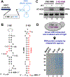
Figure 1:
Figure 1:. Analysis of proteins binding to the 3’-BE in HAC1 mRNA.
(A) The template DNA sequence of the 3’-BE- or 5’-RD RNA was placed under the control of the constitutive ADH1 promoter. The predicted structure of the RMB is shown. (B) The predicted secondary structure of 3’-BE and 5’-RD RNAs is shown. The conserved RNA motif within the 3’-BE is shown in red. The 5’-RD consisting of 5’-UTR and intron are shown in black and red, respectively. The numbers indicate the nucleotide positions. (C) To analyze mini RNA expression, cDNA was synthesized from total RNA isolated from yeast cells expressing the 3’-BE- and 5’-RD-RMB mini RNA constructs using gene-specific primers in the presence and absence of reverse transcriptase (RT). Representative of 2 independent experiments. (D) Proteins bound to the 3’-BE- or 5’-RD-RMB mini RNA were electrophoresed and stained. Representative of 1 experiment.
Figure 2:
Figure 2:. The pal1Δ pal2Δ strain is deficient in ER stress responses.
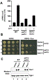
(A) & (B) The indicated yeast strains were grown, serially diluted and spotted on YEPD (A) or YEPG (B) solid medium containing the indicated concentrations of tunicamycin. Representative of 2 independent experiments. (C) The indicated yeast strains were grown in YEPG medium with or without tunicamycin (Tun). cDNA was prepared from total RNA (lower panel) and the spliced (HAC1s) and un-spliced (HAC1u) forms of HAC1 mRNA were detected. Representative of 2 independent experiments. (D) The indicated yeast strains were grown in YEPG medium with or without tunicamycin (Tun). Whole cell extracts (WCEs) were isolated and subjected to Western blot analysis using antibodies to detect Hac1 and Pgk1. Representative of 2 independent experiments.
Figure 3:
Figure 3:. Analysis of Hac1 protein expression from the intron-less HAC1 mRNA.
(A) Yeast strains (WT, kin1Δkin2Δ or pal1Δpal2Δ) transformed with a plasmid carrying the UPRE-driven LacZ reporter gene were grown in the presence (+) and absence (–) of DTT. β-galactosidase activity was measured in WCEs. Representative of 3 independent experiments. (B) The indicated yeast strains expressing vector plasmid or the same vector containing an intron-less HAC1 gene (HAC1c) were grown serially diluted and spotted on YEPG medium with or without tunicamycin. Representative of 2 independent experiments. (C) The same yeast strains as in (B) were grown in YEPG medium and whole cell extracts were isolated and subjected to Western blot analysis using antibodies to detect Hac1 and Pgk1. Representative of 2 independent experiments.
Figure 4:
Figure 4:. Pal1 and Pal2 are associated with HAC1 mRNA
(A) The pal1Δpal2Δ strains expressing WT or mutant Pal2 were tested for growth on the complete synthetic (SC) medium without uracil or the same medium containing tunicamycin. Representative of 2 independent experiments. The right panel shows the schematic representations of WT and Flag-tagged (red circle) Pal2 constructs. The intronic region of the WT PAL2 is colored light blue. The regions encoding the homologous Pal1 domains (PD, residues 125 to 272) are indicated by dark blue boxes. (B) WCEs were prepared from the same yeast strains as in (A) and subjected to Western blot analysis to detect Flag. “*” indicates Pal2. Representative of 2 independent experiments. (C) The pal1Δpal2Δ strains expressing WT Pal2 or its derivatives were tested for growth on the complete synthetic (SC) medium without uracil (Ura) or the same medium containing tunicamycin. Representative of 2 independent experiments. The right panel shows the schematic representations of WT and mutant Pal1 constructs. (D) Electrophoretic mobility shift assay (EMSA) using a fluorescein-tagged synthetic RNA corresponding to the 3’-BE (Flc-3’-BE) and recombinant His6-Pal1, His6-Pal2, His6-eIF2α or His6-Ypt1. The reaction mixture was resolved with native gels and illuminated with ultraviolet (UV)-light to see the RNA-protein interaction. Representative of 2 independent experiments. (E) Flc-3’-BE (2nd panel) was mixed with the recombinant His6-Pal2 protein (bottom panel) in a binding buffer with (lane 2) or without (lane 1) the non-fluorescent 3’-BE (3rd panel). The reaction mixture was resolved with native gels and illuminated with ultraviolet (UV)-light to see the Flc-3’-BE•Pal2 interaction. The portion of gel showing the Flc-3’-BE bound to Pal2 is shown (top panel). Representative of 2 independent experiments.
Figure 5:
Figure 5:. Kin1 phosphorylates Pal2 in vitro
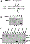
(A) The consensus sequence for Kin phosphorylation and the corresponding sequences in Pal1 and Pal2. x indicates any amino acid and pT indicates phospho-threonine. (B) The indicated proteins were purified from yeast and subjected to SDS-PAGE analysis. “*” denotes the protein of interest. Representative of 2 independent experiments. (C) Purified proteins were mixed with Kin1 in a reaction mixture containing radiolabeled ATP (γ−33P-ATP) and separated by SDS-PAGE. Radiolabel incorporation was detected by autoradiograph. Representative of 2 independent experiments.
Figure 6:
Figure 6:. Kin2 phosphorylates the residue Ser-222 of Pal2 in vitro
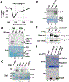
(A) Circular dichroism (CD) spectra of recombinant Pal2 (0.5 mg/mL) was collected in the wavelength range of 190–360 nm. Representative of 2 independent experiments. (B) Recombinant His6-Pal2 (residues 65–366) or eIF2α protein was incubated with purified Kin2 or PKR in a kinase reaction containing γ−32P-ATP and resolved by SDS-PAGE. Gel was stained with Coomassie blue (lower panel) and subjected to autoradiography (upper panel). Representative of 2 independent experiments. (C) Recombinant His6-Pal2 (residues 65–366) and the indicated mutants were subjected to kinase assay. Pal2-4Ala = S222A,T223A,T224A and T225A. Pal2-3Ala = T223A,T224A and T225A. Pal2-2Ala = S222A and T224A. Protein markers (kDa) are indicated. Representative of 2 independent experiments. (D) Recombinant His6-Pal1 (residues 165–499) protein was subjected to kinase assay with purified Kin2 and γ−33P-ATP as described in (B). Representative of 2 independent experiments. (E) WCEs were prepared from the indicated yeast strains containing a vector plasmid (null) or the same plasmid containing the Flag-tagged WT or 4Ala mutant Pal2. WCEs were subjected to normal SDS-PAGE or Phos-tag SDS-PAGE gel followed by Western blot analysis to detect Flag. Representative of 3 independent experiments. (F) Recombinant His6-Pal2 (residues 65–366) was subjected to kinase assay with Flag-tagged Kin2 purified from yeast (ScFlag-Kin2), GST-Kin2 purified fom E. coli (EcGST-Kin2) or GST-Kin2-T281E purified fom E. coli (EcGST-Kin2-T281E) as described in (B). Representative of 2 independent experiments.
Figure 7:
Figure 7:. Phosphorylation of Pal2 is important for ER stress responses
(A) The pal1Δ pal2Δ strains expressing WT or mutant Pal2 were tested for growth on synthetic complete medium with and without tunicamycin. Representative of 3 independent experiments. (B) WCEs from the same strains as in (A) were subjected to Western blot analysis to detect Flag. Representative of 2 independent experiments. (C) The selected pal1Δ pal2Δ yeast strains as in (A) were grown in synthetic complete medium without uracil in the presence (+) or absence (–) of tunicamycin and WCEs were subjected to Western blot analysis for Hac1 and Pgk1. Representative of 2 independent experiments. (D) The kin1Δ kin2Δ strains expressing WT Kin2, WT Pal2 or Pal2ΔN65-STTT222–225EEEE (STTT222–225EEEE) were tested for growth on synthetic complete medium with and without tunicamycin. Representative of 2 independent experiments. (E) The homologous Pal1 domains (PD) in Pal1 and Pal2 are indicated by dark blue boxes. The phosphorylation loop of Pal2 is shown.
Figure 8:
Figure 8:. Proposed role of Pal2 in the Ire1-HAC1 mediated UPR.
The ER-resident chaperone Kar2 binds to the lumenal domain of Ire1 and keeps it in an inactive form. During the ER stress, unfolded proteins (scribbled red line) accumulate inside the ER lumen and titrate Kar2, thus activating the cytoplasmic kinase and RNase domain of Ire1. The active RNase domain cleaves the intron from the HAC1 mRNA. The exons (grey bars) of HAC1 mRNA are separated by an intron (orange dashed line) that interacts with the 5’-UTR (solid black) line of mRNA to form an inhibitory RNA duplex (RD). The KA1 domain (light blue bar) binds to the KD (dark blue bar) of Kin1 or Kin2, thus keeping the KD in an inactive form. Under stress conditions, the KD is activated by phosphorylation (“P” in a red circle) within the activation loop on residue Thr281 in a trans mechanism. The active KD then phosphorylates Pal2, which is likely associated with the 3’-BE and 3’-BE-specific RNP, thus targeting HAC1 mRNA to the ER stress site.

Similar articles

Cited by

References

    1. Schroder M (2008) Endoplasmic reticulum stress responses. Cell Mol Life Sci 65, 862–894 - PMC - PubMed
    1. Walter P, and Ron D (2011) The Unfolded Protein Response: From Stress Pathway to Homeostatic Regulation. Science 334, 1081–1086 - PubMed
    1. Ron D, and Walter P (2007) Signal integration in the endoplasmic reticulum unfolded protein response. Nat Rev Mol Cell Bio 8, 519–529 - PubMed
    1. Xu C, Bailly-Maitre B, and Reed JC (2005) Endoplasmic reticulum stress: cell life and death decisions. J Clin Invest 115, 2656–2664 - PMC - PubMed
    1. Cox JS, and Walter P (1996) A novel mechanism for regulating activity of a transcription factor that controls the unfolded protein response. Cell 87, 391–404 - PubMed

Publication types

MeSH terms