

Systemic NF-κB-mediated inflammation promotes an aging phenotype in skeletal stem/progenitor cells
- PMID: 34035186
- PMCID: PMC8202837
- DOI: 10.18632/aging.203083
Systemic NF-κB-mediated inflammation promotes an aging phenotype in skeletal stem/progenitor cells
Abstract
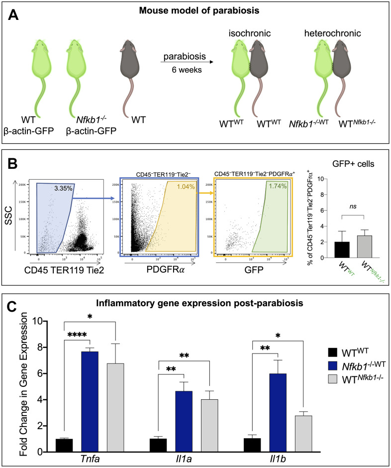
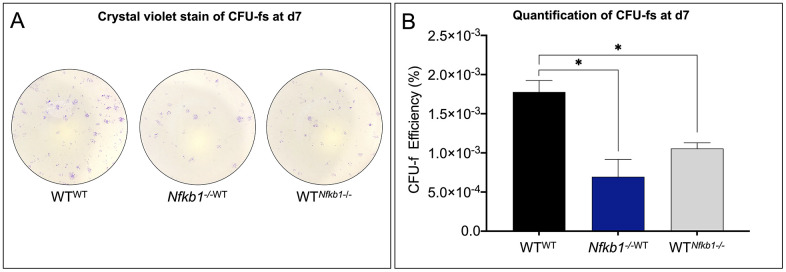
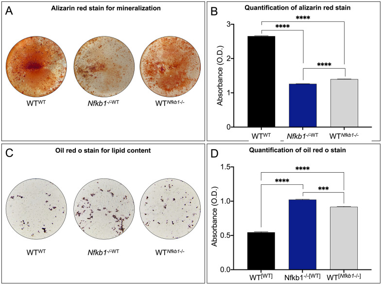
Aging tissues undergo a progressive decline in regenerative potential. This decline in regenerative responsiveness has been attributed to changes in tissue-specific stem cells and their niches. In bone, aged skeletal stem/progenitor cell dysfunction is characterized by decreased frequency and impaired osteogenic differentiation potential. This aging phenotype ultimately results in compromised regenerative responsiveness to injury. The age-associated increase of inflammatory mediators, known as inflamm-aging, has been identified as the main culprit driving skeletal stem cell dysfunction. Here, we utilized a mouse model of parabiosis to decouple aging from inflammation. Using the Nfkb1-/- mouse as a model of inflamm-aging, we demonstrate that a shared systemic circulation between a wild-type and Nfkb1-/- mouse results in an aging phenotype of the wild-type skeletal stem and progenitor cells, shown by CFU-fs and osteogenic and adipogenic differentiation assays. Our findings demonstrate that exposure to an inflammatory secretome results in a phenotype similar to the one observed in aging.
Keywords: aging; inflammation; nuclear factor kappa B; regeneration; skeletal stem cell.
Conflict of interest statement
Figures
Similar articles
-
Age-related inflammation triggers skeletal stem/progenitor cell dysfunction.Proc Natl Acad Sci U S A. 2019 Apr 2;116(14):6995-7004. doi: 10.1073/pnas.1810692116. Epub 2019 Mar 20. Proc Natl Acad Sci U S A. 2019. PMID: 30894483 Free PMC article.
-
NF-κB negatively impacts the myogenic potential of muscle-derived stem cells.Mol Ther. 2012 Mar;20(3):661-8. doi: 10.1038/mt.2011.261. Epub 2011 Dec 13. Mol Ther. 2012. PMID: 22158056 Free PMC article.
-
Inflammation rapidly recruits mammalian GMP and MDP from bone marrow into regional lymphatics.Elife. 2021 Apr 8;10:e66190. doi: 10.7554/eLife.66190. Elife. 2021. PMID: 33830019 Free PMC article.
-
Aging, metabolism and stem cells: Spotlight on muscle stem cells.Mol Cell Endocrinol. 2017 Apr 15;445:109-117. doi: 10.1016/j.mce.2016.08.021. Epub 2016 Aug 12. Mol Cell Endocrinol. 2017. PMID: 27531569 Review.
-
Bone Marrow Niches for Skeletal Progenitor Cells and their Inhabitants in Health and Disease.Curr Stem Cell Res Ther. 2019;14(4):305-319. doi: 10.2174/1574888X14666190123161447. Curr Stem Cell Res Ther. 2019. PMID: 30674266 Review.
Cited by
-
Addressing osteoblast senescence: Molecular pathways and the frontier of anti-ageing treatments.Clin Transl Med. 2025 Jul;15(7):e70417. doi: 10.1002/ctm2.70417. Clin Transl Med. 2025. PMID: 40681473 Free PMC article. Review.
-
Inflammatory Processes Affecting Bone Health and Repair.Curr Osteoporos Rep. 2023 Dec;21(6):842-853. doi: 10.1007/s11914-023-00824-4. Epub 2023 Sep 28. Curr Osteoporos Rep. 2023. PMID: 37759135 Free PMC article. Review.
-
The impact of age and sex on the inflammatory response during bone fracture healing.JBMR Plus. 2024 Feb 22;8(5):ziae023. doi: 10.1093/jbmrpl/ziae023. eCollection 2024 May. JBMR Plus. 2024. PMID: 38560342 Free PMC article.
-
Exploring Quercetin Anti-Osteoporosis Pharmacological Mechanisms with In Silico and In Vivo Models.Life (Basel). 2022 Jun 29;12(7):980. doi: 10.3390/life12070980. Life (Basel). 2022. PMID: 35888070 Free PMC article.
-
Bone regeneration in inflammation with aging and cell-based immunomodulatory therapy.Inflamm Regen. 2023 May 25;43(1):29. doi: 10.1186/s41232-023-00279-1. Inflamm Regen. 2023. PMID: 37231450 Free PMC article. Review.
References
-
- Laschober GT, Brunauer R, Jamnig A, Singh S, Hafen U, Fehrer C, Kloss F, Gassner R, Lepperdinger G. Age-specific changes of mesenchymal stem cells are paralleled by upregulation of CD106 expression as a response to an inflammatory environment. Rejuvenation Res. 2011; 14:119–31. 10.1089/rej.2010.1077 - DOI - PubMed
Publication types
MeSH terms
Substances
Grants and funding
LinkOut - more resources
Full Text Sources
Other Literature Sources
Medical
Miscellaneous
