. 2021 May 26;6(1):187.
doi: 10.1038/s41392-021-00579-3.

Exosomal S100A4 derived from highly metastatic hepatocellular carcinoma cells promotes metastasis by activating STAT3

Affiliations

Exosomal S100A4 derived from highly metastatic hepatocellular carcinoma cells promotes metastasis by activating STAT3

Haoting Sun et al. Signal Transduct Target Ther. .

Abstract

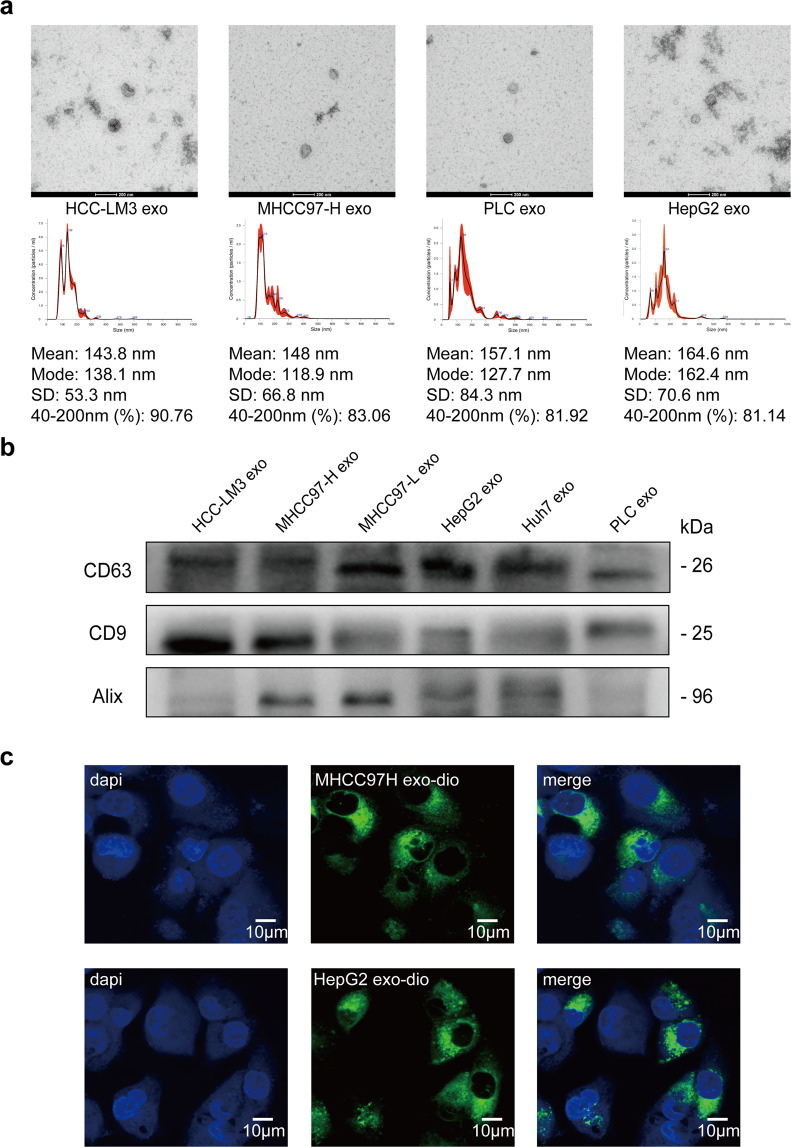
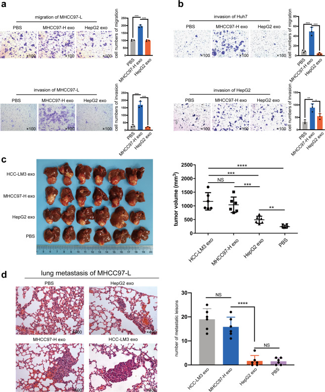
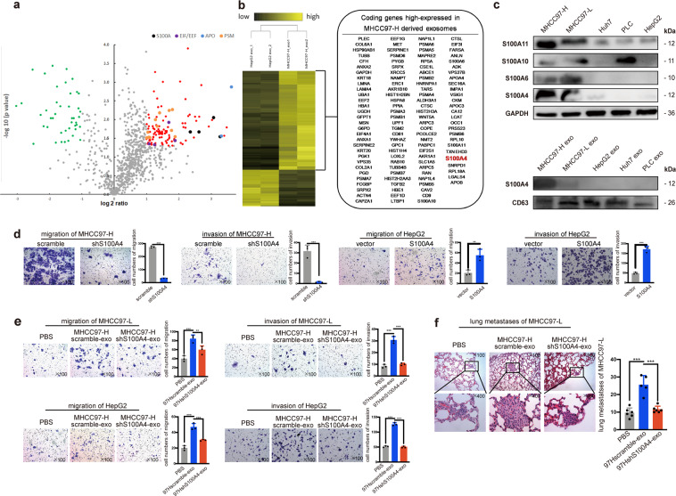
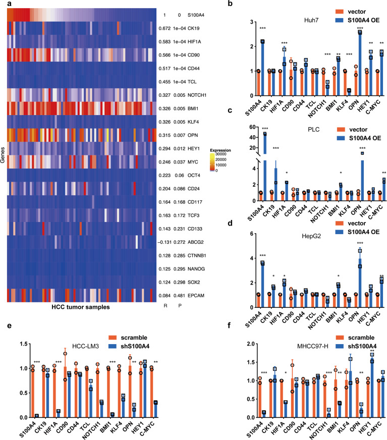
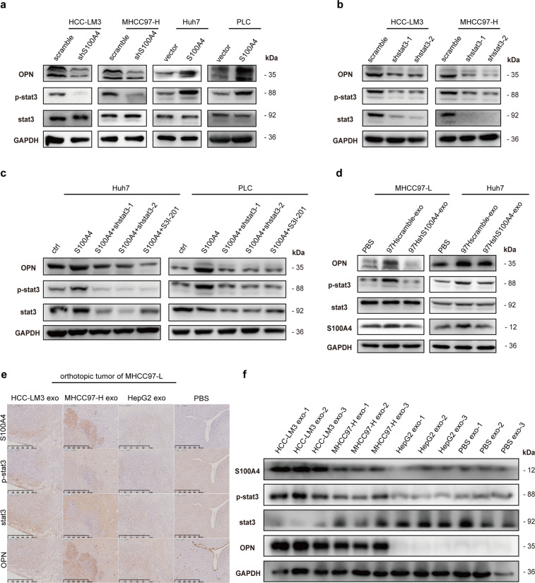
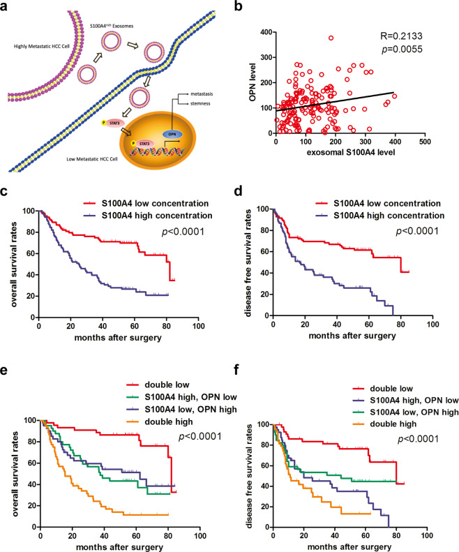
Intercellular cross-talk plays important roles in cancer progression and metastasis. Yet how these cancer cells interact with each other is still largely unknown. Exosomes released by tumor cells have been proved to be effective cell-to-cell signal mediators. We explored the functional roles of exosomes in metastasis and the potential prognostic values for hepatocellular carcinoma (HCC). Exosomes were extracted from HCC cells of different metastatic potentials. The metastatic effects of exosomes derived from highly metastatic HCC cells (HMH) were evaluated both in vitro and in vivo. Exosomal proteins were identified with iTRAQ mass spectrum and verified in cell lines, xenograft tumor samples, and functional analyses. Exosomes released by HMH significantly enhanced the in vitro invasion and in vivo metastasis of low metastatic HCC cells (LMH). S100 calcium-binding protein A4 (S100A4) was identified as a functional factor in exosomes derived from HMH. S100A4rich exosomes significantly promoted tumor metastasis both in vitro and in vivo compared with S100A4low exosomes or controls. Moreover, exosomal S100A4 could induce expression of osteopontin (OPN), along with other tumor metastasis/stemness-related genes. Exosomal S100A4 activated OPN transcription via STAT3 phosphorylation. HCC patients with high exosomal S100A4 in plasma also had a poorer prognosis. In conclusion, exosomes from HMH could promote the metastatic potential of LMH, and exosomal S100A4 is a key enhancer for HCC metastasis, activating STAT3 phosphorylation and up-regulating OPN expression. This suggested exosomal S100A4 to be a novel prognostic marker and therapeutic target for HCC metastasis.


Conflict of interest statement

The authors declare no competing interests.

Figures

Fig. 1
Fig. 1
Isolation and identification of exosomes released and up-taken by HCC cells. a Transmission electron microscopy of isolated exosomes from HCC cells (HCC-LM3, MHCC97-H, PLC, and HepG2, Scale bar: 200 nm). The concentration and size distribution were determined by nanoparticle tracking analysis (NTA). b Western blot of exosomal marker CD63, CD9, and Alix in exosomes isolated from HCC cell lines (HCC-LM3, MHCC97-H, MHCC-97-L, HepG2, Huh7, and PLC). c Laser scanning confocal microscope images of DIO treated exosomes (MHCC97H exo and HepG2 exo) up-taken by low metastatic HCC cells (MHCC97-L)
Fig. 2
Fig. 2
Highly metastatic HCC cells (HMH) derived exosomes enhance metastatic potential of low metastatic HCC cells (LHM) both in vitro and in vivo. a, b Assessment of migration and invasion in vitro. LHM (MHCC97-L, Huh7, and HepG2) pre-treated with MHCC97-H exosomes, HepG2 exosomes, or PBS (as negative control) for 24 h. The migration ability of MHCC97-L and invasion ability of all the three cell lines were significantly enhanced by MHCC97-H exosomes compared to the control groups. c Orthotopic implantation liver tumors from nude mice. MHCC97-L cells treated with HMH exosomes (HCC-LM3 exosomes or MHCC97-H exosomes) formed larger liver tumors. d Lung metastasis of mice injected with MHCC97-L. MHCC97-L cells treated with HMH exosomes formed significantly more lung metastatic lesions. All the in vitro assays were conducted three times with three repetitions. Error bars represent the mean ± SD, and the dots represent the value of repetitions in one experiment; *P < 0.05, **P < 0.01, ***P < 0.001, ****P < 0.0001, ns: no significance. An unpaired t test was employed in (a) and (b), one-way ANOVA followed by Bonferroni’s post hoc test was employed in (c) and (d)
Fig. 3
Fig. 3
Exosomal S100A4 is a key promoter to enhance metastatic potential. a iTRAQ mass spectrum screening was performed to analyze the protein cargo of exosomes derived from MHCC97-H (HMH-exosomes) and HepG2 exosomes (LMH-exosomes). Proteins were clustered at the 2-fold changes with a p value less than 0.05. The volcano plots revealed a total 116 up-regulated proteins (right quadrant, generally in red, S100A family in black, EIF/EEF family in purple, APO family in blue and PSM family in orange), and yet 43 proteins were found down-regulated (left quadrant, in green). b The results are also expressed as a heat map, from which 116 up-regulated proteins are shown. c Diverse expression abundance of S100A protein family in HCC cell lines. Western blot showed that expressions of S100A4 were the highest in MHCC97-H among HCC cell lines. S100A4 was only highly expressed in exosomes derived from MHCC97-H. d S100A4 knock-down in MHCC97-H results in inhibited migration and invasion ability. Overexpression of S100A4 enhanced migration and invasion in HepG2. e S100A4rich exosomes significantly promotes migration and invasive abilities of cells. Exosomal S100A4 promotes the migration and invasive abilities of cells in vitro. Exosomes of MHCC97-H cells with S100A4 knockdown were defined as S100A4low exosomes, ones derived from the scrambled counterparts were defined as S100A4rich exosomes. S100A4rich exosomes were used to treat MHCC97-L and HepG2 cells for 24 h before migration and invasion assays while PBS and S100A4low exosomes were used as negative controls. f Exosomal S100A4 promotes the metastatic potential in vivo. MHCC97-L cells treated with PBS, S100A4rich exosomes or S100A4low exosomes were injected in nude mice to form lung metastasis model. All the in vitro assays were conducted three times with three repetitions. Error bars represent the mean ± SD, and the dots represent the value of each experiment; *P < 0.05, **P < 0.01, ***P < 0.001, ****P < 0.0001, ns: no significance. Statistical significance was determined by unpaired t test
Fig. 4
Fig. 4
S100A4 expression positively correlates with OPN. a Correlation between S100A4 and stemness-related genes. The GEO datasheet GSE39791 were used for correlation analyzes. Among the selected genes, 11 genes including OPN showed significant positive correlation with S100A4. Real-time PCR analyzes revealed the change of mRNA expression after S100A4 overexpression in low metastatic HCC cell lines, Huh7 (b), PLC (c), and HepG2 (d). In highly metastatic HCC cell lines, HCC-LM3 (d) and MHCC97-H (f), with S100A4 knockdown. Only OPN and its downstream genes (HIF1 and BMI1) in accordance with the tendency of S100A4 expression after knockdown and overexpression. Real-time PCR was conducted three times with two repetitions, and the dots represent the value of repetitions in one experiment. Error bars represent the mean ± SD; *P < 0.05, **P < 0.01, ***P < 0.001, ****P < 0.0001
Fig. 5
Fig. 5
Exosomal S100A4 activates OPN transcription via stat3 phosphorylation. a Western blot indicated that OPN and stat3 phosphorylation (p-stat3) levels follow the change of S100A4 level, while stat3 level maintains stable. b Western blot showed that knockdown of stat3 expression in high metastatic cell lines (HCC-LM3 and MCC97-H) spontaneously inhibits p-stat3 and OPN expressions. c Overexpression of stat3 in low metastatic cell lines (Huh7 and PLC) enhanced p-stat3 and OPN level, while such effect alleviated by stat3 knock down or stat3 inhibitor S3I-201, suggesting a relative up-stream regulation of p-stat3 to OPN. d Western blot showed that stat3 phosphorylation and OPN are significantly enhanced after S100A4rich exosomes (97Hshscramble-exo) treatment. e, f Immunohistochemical staining and Western blot quantification of orthotopic tumors from mice showed that HMH exosomes (HCC-LM3 exosomes and MHCC97-H exosomes) promote stat3 phosphorylation and OPN expression in tumor tissue compared to PBS and LMH exosomes (HepG2 exosomes). Western blot was conducted three times
Fig. 6
Fig. 6
Plasma exosomal S100A4 and OPN levels jointly serve as a powerful postoperative prognostic factor for HCC patients. a Working model of exosomal S100A4 promoting HCC metastasis. S100A4rich exosomes released by highly metastatic HCC cells enhanced metastatic potential of low metastatic HCC cells via STAT3 phosphorylation and OPN up-regulation. b Correlation analysis on plasma OPN level and plasma exosomal level of S100A4 showed positive correlation in clinical samples. c, d Patients with low plasma exosomal S100A4 level had significantly better overall survival (OS) and time to treat (TTR) compared with HCC patients with high plasma exosomal S100A4 level. e, f Patients with both low plasma exosomal S100A4 level and OPN level had the longest OS and DFS among the four subgroups, which were divided according to combination of exosomal S100A4 and OPN. For each cohort, different subgroups were plotted according to the cut-off values of exosomal S100A4 (98.54 pg/ml) and OPN (103.78 ng/ml). Error bars represent the mean ± SD, and the dots represent the value of each experiment; *P < 0.05, **P < 0.01, ***P < 0.001, ****P < 0.0001, ns: no significance. Simple linear regression was employed in (b), log-rank test was employed in (cf)

References

    1. Gupta GP, Massague J. Cancer metastasis: building a framework. Cell. 2006;127:679–695. doi: 10.1016/j.cell.2006.11.001. - DOI - PubMed
    1. Hanahan D, Weinberg RA. Hallmarks of cancer: the next generation. Cell. 2011;144:646–674. doi: 10.1016/j.cell.2011.02.013. - DOI - PubMed
    1. Villanueva A. Hepatocellular carcinoma. N. Engl. J. Med. 2019;380:1450–1462. doi: 10.1056/NEJMra1713263. - DOI - PubMed
    1. Liu J, Dang H, Wang XW. The significance of intertumor and intratumor heterogeneity in liver cancer. Exp. Mol. Med. 2018;50:e416. doi: 10.1038/emm.2017.165. - DOI - PMC - PubMed
    1. Becker A, et al. Extracellular vesicles in cancer: cell-to-cell mediators of metastasis. Cancer Cell. 2016;30:836–848. doi: 10.1016/j.ccell.2016.10.009. - DOI - PMC - PubMed

Publication types

MeSH terms