

doi: 10.1038/s41421-021-00264-3.
Structural basis for neutralization of an anicteric hepatitis associated echovirus by a potent neutralizing antibody
Affiliations
- PMID: 34035235
- PMCID: PMC8149713
- DOI: 10.1038/s41421-021-00264-3
Item in Clipboard
Structural basis for neutralization of an anicteric hepatitis associated echovirus by a potent neutralizing antibody
Cell Discov.
.
No abstract available
Conflict of interest statement
The authors declare no competing interests.
Figures
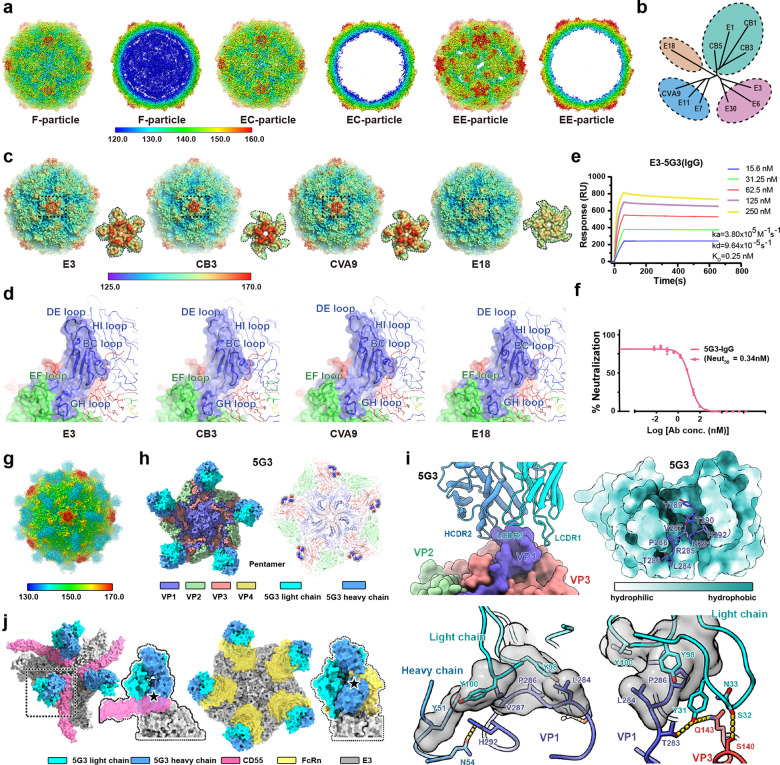
a Surface representations of E3 F-, EC-, and EE-particles and thin slices of the corresponding central sections viewed along the two-fold axes, respectively. The surface of the virus is colored by rainbow-color based on the distance of the viral elements from the center, starting with blue (closest) through green, yellow, and orange to red (farthest from the center). b Structure-based evolutionary relationship among the representative viruses from HEV-Bs: E18, echovirus 18; E3, echovirus 3; CVB1, coxsackievirus B1; E6, echovirus 6; E30, echovirus 30; CVB5, coxsackievirus B5; CVB3, coxsackievirus B3; E1, echovirus 1; CVA9, coxsackievirus A9; E11, echovirus 11. c Comparisons of the surface of E3 F-particle with those of other representative members of HEV-B (E18 (PDB: 6HBG), CVB3 (PDB: 4GB3), and CVA9 (PDB: 1D4M)). The color scheme is the same as in a. The “mesa” located at the top is marked using a black dotted square for each particle. d Structural details of the protomer (VP1: blue; VP2: green; VP3: red) of representative particles. The loops surrounding the canyon walls (VP1 BC loop, VP1 GH loop, and VP2 EF loop) and the loops surrounding the “mesa” structures are labeled in corresponding colors. e The binding affinities of E3 F-particle to 5G3 IgG estimated by SPR. f Neutralization of E3 by 5G3 using plaque-reduction neutralization test (PRNT). The Neut50 value of 5G3 was 0.34 nM. g Surface representation of E3-5G3 complex. The viral capsid is colored the same as in a, and the 5G3 Fab is colored in cyan. h 5G3 Fab occupancy (left) and epitopes (right) located on viral pentamer. The pentamer is shown as surface (left) and cartoon (right), respectively. Residues comprising 5G3 epitope are shown as spheres. i The E3–5G3 binding interface. 5G3 are shown as cartoon, while E3 is shown as surface. The residues of the VP1 C-terminal (blue) insert into the 5G3 Fab hydrophobic pocket. The change of color reflects hydrophilic or hydrophobic nature of the residues, ranging from white (hydrophilic) to cyan (hydrophobic). Residues involved in the interactions between the virus (VP1: blue, VP3: red) and 5G3 are shown as sticks, and the hydrophobic interactions are shown as surface and colored in grey. Hydrogen bonds are marked as yellow dashes. j Clashes between 5G3 and two E3 receptors, CD55 (left)/FcRn (right). The pentamer, 5G3 light chain, 5G3 heavy chain, receptor CD55, and receptor FcRn are colored in grey, cyan, blue, magenta, and yellow, respectively.
Similar articles
-
Neutralization of different echovirus serotypes by individual lots of intravenous immunoglobulin.J Med Virol. 2011 Feb;83(2):305-10. doi: 10.1002/jmv.21980. J Med Virol. 2011. PMID: 21181927
-
Echovirus 3 infection associated with anicteric hepatitis.Am J Dis Child. 1982 Aug;136(8):744-5. doi: 10.1001/archpedi.1982.03970440088028. Am J Dis Child. 1982. PMID: 7102628 No abstract available.
-
Seroepidemiology of echovirus 30 in Korean children.World J Pediatr. 2017 Dec;13(6):611-614. doi: 10.1007/s12519-017-0058-x. Epub 2017 Aug 2. World J Pediatr. 2017. PMID: 28766163
-
Defining Breadth of Hepatitis C Virus Neutralization.Front Immunol. 2018 Aug 2;9:1703. doi: 10.3389/fimmu.2018.01703. eCollection 2018. Front Immunol. 2018. PMID: 30116237 Free PMC article. Review.
-
Neutralizing antibodies in hepatitis C virus infection.World J Gastroenterol. 2007 Sep 28;13(36):4824-30. doi: 10.3748/wjg.v13.i36.4824. World J Gastroenterol. 2007. PMID: 17828813 Free PMC article. Review.
Cited by
-
Atomic Structures of Coxsackievirus B5 Provide Key Information on Viral Evolution and Survival.J Virol. 2022 May 11;96(9):e0010522. doi: 10.1128/jvi.00105-22. Epub 2022 Apr 20. J Virol. 2022. PMID: 35442060 Free PMC article.
-
Immunogenicity assessment of Hepatitis A-VP1 and Hepatitis B surface antigen (HBsAg) fusion protein: a novel bivalent vaccine candidate.Iran J Microbiol. 2025 Aug;17(4):636-643. doi: 10.18502/ijm.v17i4.19257. Iran J Microbiol. 2025. PMID: 40785716 Free PMC article.
-
Structural Basis for the Immunogenicity of the C-Terminus of VP1 of Echovirus 3 Revealed by the Binding of a Neutralizing Antibody.Viruses. 2022 Oct 22;14(11):2322. doi: 10.3390/v14112322. Viruses. 2022. PMID: 36366420 Free PMC article.
References
-
- Leggiadro RJ, Chwatsky DN, Zucker SW. Echovirus 3 infection associated with anicteric hepatitis. Am. J. Dis. Child. 1982;136:744–745. - PubMed
Publication types
LinkOut - more resources
Full Text Sources
Other Literature Sources
