. 2021 May 25;12(1):3091.
doi: 10.1038/s41467-021-23408-1.

The P-type ATPase transporter ATP7A promotes angiogenesis by limiting autophagic degradation of VEGFR2

Affiliations

The P-type ATPase transporter ATP7A promotes angiogenesis by limiting autophagic degradation of VEGFR2

Dipankar Ash et al. Nat Commun. .

Abstract

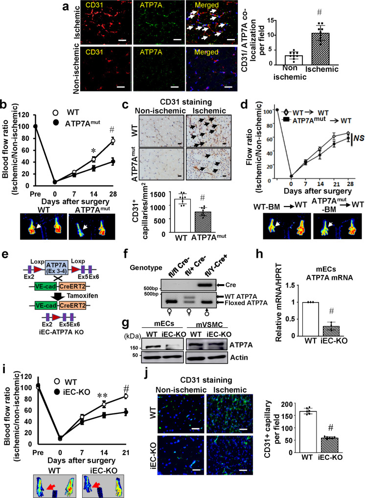
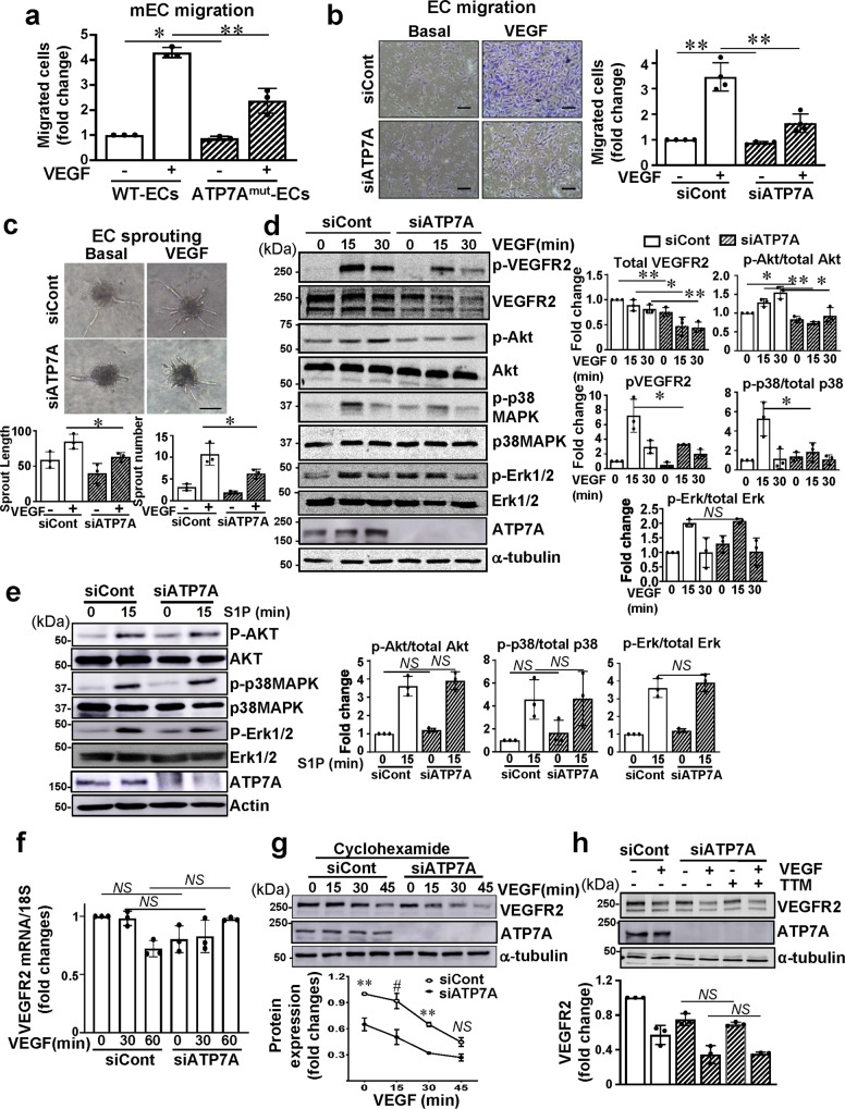
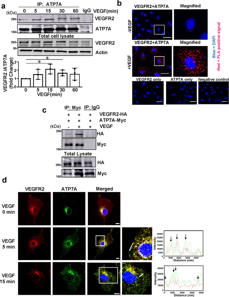
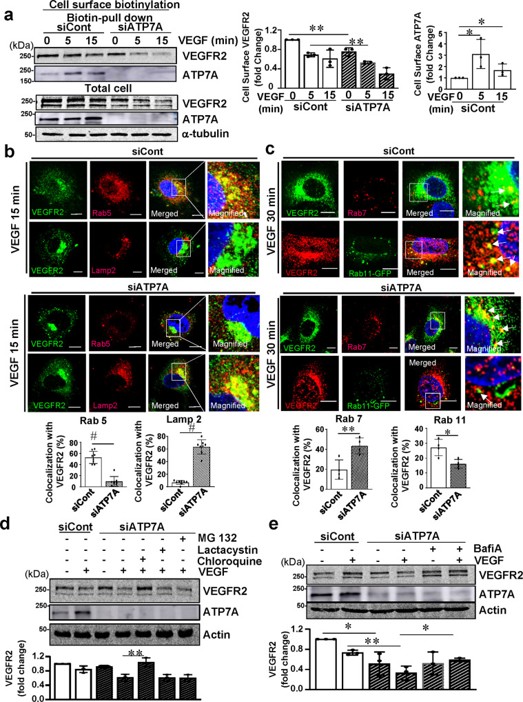
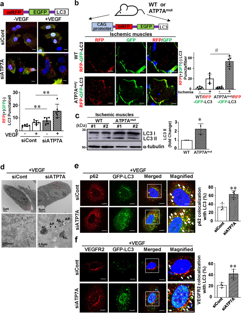
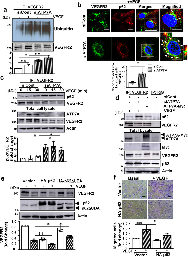
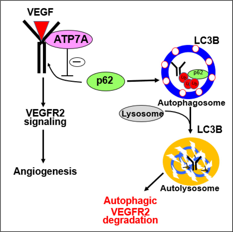
VEGFR2 (KDR/Flk1) signaling in endothelial cells (ECs) plays a central role in angiogenesis. The P-type ATPase transporter ATP7A regulates copper homeostasis, and its role in VEGFR2 signaling and angiogenesis is entirely unknown. Here, we describe the unexpected crosstalk between the Copper transporter ATP7A, autophagy, and VEGFR2 degradation. The functional significance of this Copper transporter was demonstrated by the finding that inducible EC-specific ATP7A deficient mice or ATP7A-dysfunctional ATP7Amut mice showed impaired post-ischemic neovascularization. In ECs, loss of ATP7A inhibited VEGF-induced VEGFR2 signaling and angiogenic responses, in part by promoting ligand-induced VEGFR2 protein degradation. Mechanistically, VEGF stimulated ATP7A translocation from the trans-Golgi network to the plasma membrane where it bound to VEGFR2, which prevented autophagy-mediated lysosomal VEGFR2 degradation by inhibiting autophagic cargo/adapter p62/SQSTM1 binding to ubiquitinated VEGFR2. Enhanced autophagy flux due to ATP7A dysfunction in vivo was confirmed by autophagy reporter CAG-ATP7Amut -RFP-EGFP-LC3 transgenic mice. In summary, our study uncovers a novel function of ATP7A to limit autophagy-mediated degradation of VEGFR2, thereby promoting VEGFR2 signaling and angiogenesis, which restores perfusion recovery and neovascularization. Thus, endothelial ATP7A is identified as a potential therapeutic target for treatment of ischemic cardiovascular diseases.


Conflict of interest statement

The authors declare no competing interests.

Figures

Fig. 1
Fig. 1. Endothelial ATP7A is required for ischemia-induced neovascularization.
a CD31 (red, Endothelial Cell (EC) marker) or ATP7A (green) staining or their colocalization (merged, white arrows) in ischemic and non-ischemic gastrocnemius muscles at day 14 post-surgery. Scale bars = 50 μm. The bar graph represents CD31-ATP7A co-localized cell numbers per field. n = 6, #p < 0.0001 (two-tailed unpaired t-test). b Blood flow recovery as determined by the ratio of foot perfusion between ischemic (left) and ischemic (right) legs after hindlimb ischemia in Wild Type (WT) and ATP7Amut mice. n = 5, *p < 0.05, #p < 0.0001 (two-way ANOVA followed by Bonferroni’s multiple comparison analysis). Bottom panels show representative laser Doppler images (red arrows show ischemic foot) of legs at day 28 after ischemia in WT and ATP7Amut mice. c Capillary density (CD31 staining) in ischemic and non-ischemic muscles of WT and ATP7Amut mice at day 14. Scale bars = 50 μm. Bottom panels show their quantitative analysis (number of capillaries per mm square, n = 8, #p < 0.0001 (two-tailed unpaired t-test). d Bone marrow (BM) from WT or ATP7Amut mice were transplanted to irradiated WT mice. After 6 weeks, mice were subjected to hindlimb ischemia and limb blood flow was measured. n = 6, NS = non-significant (two-way ANOVA followed by Bonferroni’s multiple comparison analysis). The bottom panels show representative laser Doppler images on day 28. e Schematic representation generating inducible EC-specific hemizygous male ATP7A knockout (iEC-ATP7AKO) mice by crossing homozygous floxed females (ATP7Afl/fl) with mice expressing the Cre recombinase located downstream of tamoxifen-inducible VE-Cadherin promotor (ATP7A+/Y; Cdh5-CreERT2+/−). f Representative gel images of genotyping from ATP7Afl/flCre (female), ATP7Afl/+Cre (female) and ATP7Afl/Y Cre+ (male) mice. A similar pair of gels were resolved to genotype an average of 50 mice/breeding pair with n = 3 independent breeding pairs. g, h ATP7A protein (g) and mRNA (h) expression in ECs and aortic vascular smooth muscle cells isolated from WT and iEC-KO mice. n = 3 animals/group, #p = 0.0005 (two-tailed unpaired t-test). i Blood flow recovery after hindlimb ischemia in WT and iEC-KO mice. The bottom panels show representative laser Doppler images on day 21. n = 5 animals/group, **p = 0.01, #p < 0.0001 (two-way ANOVA followed by Bonferroni’s multiple comparison analysis). j Capillary density (CD31 staining) in ischemic and non-ischemic muscles of WT and iEC-KO mice at day 14. Right panels show quantification for the number of capillaries per field in ischemic muscles. n = 6, #p < 0.0001 (two-tailed unpaired t-test). Scale bars = 50 μm. Data are mean ± SEM.
Fig. 2
Fig. 2. ATP7A knockdown inhibits VEGFR2 signaling via promoting VEGFR2 degradation in a Cu-independent manner.
a, b Endothelial cell (EC) migration measured using modified Boyden chamber method in ECs isolated from Wild Type (WT) and ATP7Amut mice (a) or human umbilical vein endothelial cells (HUVECs) transfected with control or ATP7A siRNAs (b) stimulated with vascular endothelial growth factor (VEGF) (20 ng/ml) for 6 h. a n = 3, *p = 0.0485, **p = 0.0034; b n = 3, **p = 0.0029, **p = 0.0016 (two-tailed unpaired t-test). Scale bars = 100 μm. c EC spheroid spouting assay in HUVECs transfected with control or ATP7A siRNAs. Right panels show an average number of sprouts and tube length per field. Scale bars = 150 μm. n = 3, *p = 0.038, *p = 0.0469 (two-tailed unpaired t-test). d, e HUVECs transfected with control or ATP7A siRNAs were stimulated with VEGF (20 ng/ml) or sphingosine-1-phosphate (S1P) (10 µM) for the indicated time. Lysates were immunoblotted (IB) with indicated antibodies (Abs). α-tubulin as a loading control. The graph represents the averaged fold change over the basal control. d n = 3, VEGFR2 **p = 0.0086, *p = 0.0272, **p = 0.0092; pAKT/total AKT: *p = 0.0279, **p = 0.0011, *p = 0.0177; p-VEGFR2: *p = 0.0393; p-p38/total p38: *p = 0.0413; NS = non significant; e: n = 3, NS = non significant (two-tailed unpaired t-test). f. HUVECs transfected with control or ATP7A siRNAs were stimulated with VEGF (20 ng/ml) and mRNA level for VEGFR2 normalized by 18S was measured. n = 3, NS = non significant (two-tailed unpaired t-test). g HUVECs transfected with control or ATP7A siRNA pretreated with cyclohexamide (10 nM for 10 min) were stimulated with VEGF (20 ng/ml). Lysates were IB with anti-VEGFR2 or α-tubulin (loading control) Abs. n = 3, **p < 0.01, #p < 0.0001 (two-way ANOVA followed by Bonferroni’s multiple comparison analysis). h HUVECs transfected with control or ATP7A siRNAs were pretreated with tetrathiomolybdate (TTM) (10 nM) for 24 h, followed by VEGF stimulation for 30 min. Lysates were IB with anti-VEGFR2 or α-tubulin Abs. n = 3, NS = non significant (two-tailed unpaired t-test). Data are mean ± SEM.
Fig. 3
Fig. 3. VEGF stimulation promotes ATP7A binding with VEGFR2.
a Human umbilical vein endothelial cells (HUVECs) were stimulated with vascular endothelial growth factor (VEGF) (20 ng/ml) for the indicated time. Lysates were immunoprecipitated (IP) with anti-ATP7A antibody (Ab) followed by immunoblotting (IB) with VEGFR2 or ATP7A Ab. The graph represents the averaged fold change of the VEGFR2/ATP7A ratio over the basal ratio. n = 3, *p = 0.0275, *p = 0.0303 (two-tailed unpaired t-test). Data are mean ± SEM. b HUVECs stimulated with VEGF (20 ng/ml) for 30 min were fixed in 4% paraformaldehyde. In situ Proximity, Ligation Assay (PLA) was performed to show the interaction of ATP7A with VEGFR2. Red dots indicate PLA positive signal. Either ATP7A or VEGFR2 or no antibody was used as a negative control. The scale bar;10 µm. (n = 3). c Cos7 cells were transfected with ATP7A-myc and VEGFR2-HA with or without VEGF (20 ng/ml) stimulation. Lysates were subjected to anti-Myc IP and anti-HA IB. (n = 3). d Co-localization of VEGFR2 and ATP7A in HUVECs stimulated with VEGF (20 ng/ml), showing yellow fluorescence in the merged image, was analyzed by comparing the fluorescence intensity for each protein (white line on the enlarged image). Scale bars = 10 μm. (n = 3).
Fig. 4
Fig. 4. ATP7A knockdown enhances lysosomal degradation of VEGFR2.
a Human umbilical vein endothelial cells (HUVECs) transfected with control or ATP7A siRNAs were stimulated with vascular endothelial growth factor (VEGF) (20 ng/ml). Lysates were used for cell surface biotinylation using sulfo-NHS-SS-biotin to measure cell surface VEGFR2 or ATP7A by biotin pull-down, followed by immunoblotting (IB) with anti-VEGFR2 or ATP7A antibody (Ab). Non-immunoprecipitated (IP) total cell lysate was used for IB with Abs indicated. The bar graph represents the averaged cell-surface VEGFR2 or ATP7A protein levels over the basal control. n = 3, VEGFR2: **p = 0.0066, **p = 0.0046; ATP7A: *p = 0.047, *p = 0.0343 (two-tailed unpaired t-test). b HUVECs transfected with control or ATP7A siRNAs stimulated with VEGF (20 ng/ml) for 15 min were used for co-localization analysis for VEGFR2 and Rab5 or Lamp2. Yellow fluorescence in merged images indicates their colocalization. Scale bars = 10 μm. The bar graph represents the percentage of colocalization with VEGFR2. n = 8, Rab5: #p < 0.0001; lamp2: #p < 0.0001 (two-tailed unpaired t-test). c HUVECs transfected with control or ATP7A siRNAs and stimulated with VEGF (20 ng/ml) for 30 min were used for co-localization analysis for VEGFR2 and Rab7 using the corresponding antibodies. For VEGFR2-Rab11 colocalization, Rab11 was detected through Rab11-GFP plasmid over-expression and VEGFR2 was detected through an anti-VEGFR2 antibody. Yellow fluorescence in merged images indicates their colocalization. Scale bars = 10 μm. The bar graph represents the percentage of colocalization with VEGFR2. n = 4, Rab7: **p = 0.009; Rab11: *p = 0.0128 (two-tailed unpaired t-test). d, e HUVECs transfected with control or ATP7A siRNAs were pretreated with lysosome inhibitor chloroquine (500 µM for 30 min) or proteasome inhibitors MG132 (carbobenzoxy-Leu-Leu-leucinal) (10 µM for 30 min) for 30 min, or lactacystin (10 µM for 60 min) (d) or bafilomycin A1 (5 nM for 60 min) (e) followed by VEGF stimulation for 30 min. Lysates were IB with anti-VEGFR2 or actin loading control. d n = 3, **p = 0.009; e n = 3, *p = 0.0223, **p = 0.0027, *p = 0.0122 (two-tailed unpaired t-test). Data are mean ± SEM.
Fig. 5
Fig. 5. ATP7A depletion induces autophagy and promotes VEGFR2 colocalization with LC3.
a Immunofluorescence analysis of LC3 (Microtubule-associated protein 1 A/1B-light chain 3)-RFP and GFP puncta (mature and immature LC3 puncta, respectively), human umbilical vein endothelial cells (HUVECs) transfected with LC3-RFP-GFP plasmids in the presence of either ATP7A or control siRNAs were treated with vascular endothelial growth factor (VEGF) (20 ng/ml) for 30 min. Scale, 10 μm. The bar graph represents averaged number of RFP positive and GFP negative puncta per cell. n = 6, **p = 0.0033, **p = 0.0022 (two-tailed unpaired t-test). b. ATP7Amut mice crossed with RFP-EGFP-LC3 transgenic mice to generate ATP7Amut/RFP-EGFP-LC3 transgenic reporter mice. Wild Type (WT) and ATP7Amut reporter mice were subjected to hind limb ischemia. Gastrocnemius muscles were harvested at 3 days after ischemia and examined for RFP and GFP expression. The scale bar; 10 µm. Bar graph represents averaged number of RFP positive and GFP negative puncta per fiber. n = 5, #p = 0.0005 (two-tailed unpaired t-test). c Representative western analysis of ischemic gastrocnemius muscles of WT and ATP7Amut mice at 3 days after ischemia. The bar graph represents averaged fold change over control and tubulin as a loading control. n = 3, *p = 0.0196 (two-tailed unpaired t-test). d HUVECs transfected with control or ATP7A siRNAs were stimulated with VEGF (20 ng/ml) for 30 min and were analyzed by transmission electron microscopy. Double membrane indicates autophagosome (AP) and black vesicle included AP indicated autolysosome (AL). (n = 3). e, f HUVECs transfected with LC3-GFP plasmid in the presence of either ATP7A or control siRNAs were stimulated with VEGF (20 ng/ml) for 30 min. Cells were stained with anti p62 or VEGFR2 antibodies. The right panels depict magnified images of the boxed areas seen in the left panels. The scale bar: 10 µm. Bar graph represents averaged number of p62 or VEGFR2 and LC3 colocalized puncta per cell. e n = 4, **p = 0.0016; f n = 4 **p = 0.0036 (two-tailed unpaired t-test). Data are mean ± SEM.
Fig. 6
Fig. 6. ATP7A protects against VEGFR2 degradation by preventing VEGFR2 ubiquitination and its binding with p62.
a Human umbilical vein endothelial cells (HUVECs) transfected with control or ATP7A siRNAs were stimulated with vascular endothelial growth factor (VEGF) (20 ng/ml) for 30 min in the presence of N-ethylmaleimide (5 mM). Lysates were IP with VEGFR2 followed by immunoblotting (IB) with anti Ubiquitin antibody (Ab) to detect VEGFR2 ubiquitination. The bar graph represents averaged fold change over the control. n = 3, **p = 0.0031, #p = 0.0002, **p = 0.0023 (two-tailed unpaired t-test). b HUVECs transfected with ATP7A or control siRNAs were stimulated with VEGF (20 ng/ml) for 30 min and analyzed by immunofluorescence using VEGFR2 and p62 Abs. The scale bar = 10 µm. Bar graph represents averaged number of VEGFR2 and p62 colocalized dots per cell. n = 5, #p = 0.0007 (two-tailed unpaired t-test). c HUVECs transfected with control or ATP7A siRNAs were stimulated with VEGF (20 ng/ml) for 15 and 30 min. Lysates were immunoprecipitated (IP) with VEGFR2 Ab followed by IB with p62 or VEGFR2 Abs. n = 3 independent experiments, #p = 0.0007, *p = 0.0232, *p = 0.0294 (two-tailed unpaired t-test). d Bovine aortic endothelial cells (BAEC) transfected with bovine control or ATP7A siRNAs along with empty vector or ATP7A-Myc plasmid and cells were stimulated with VEGF (20 ng/ml) for 30 min. Lysates were IP with VEGFR2 Ab followed by IB with p62 or VEGFR2 Abs. (n = 3). e HUVECs transfected with Adenovirus expressing p62 WT or p62 lacking ubiquitin-associated (UBA) domain were treated with VEGF (20 ng/ml) for 60 min. Lysates were used for IB with VEGFR2, HA-p62. n = 3 independent experiments,*p = 0.0145, **p = 0.0082 (two-tailed unpaired t-test). f HUVECs transfected with empty vector or HA-p62 were used to measure basal and VEGF-induced endothelial cell (EC) migration using the modified Boyden chamber method. Bar graph represents averaged fold change over control. Scale bars = 100 μm. n = 3, **p = 0.0059, *p = 0.0499 (two-tailed unpaired t-test). Data are mean ± SEM.
Fig. 7
Fig. 7. Proposed model.
Proposed model showing that ATP7A binds to VEGFR2 in endothelial cells in response to VEGF, which protects against p62/SQSTM1-mediated autophagic and lysosomal degradation of VEGFR2. This in turn promotes VEGFR2 signaling, thereby promoting angiogenesis and ischemic neovascularization.

Similar articles

Cited by

References

    1. Carmeliet P, Jain RK. Molecular mechanisms and clinical applications of angiogenesis. Nature. 2011;473:298–307. doi: 10.1038/nature10144. - DOI - PMC - PubMed
    1. Simons M, Gordon E, Claesson-Welsh L. Mechanisms and regulation of endothelial VEGF receptor signalling. Nat. Rev. Mol. Cell Biol. 2016;17:611–625. doi: 10.1038/nrm.2016.87. - DOI - PubMed
    1. Koch S, Tugues S, Li X, Gualandi L, Claesson-Welsh L. Signal transduction by vascular endothelial growth factor receptors. Biochem. J. 2011;437:169–183. doi: 10.1042/BJ20110301. - DOI - PubMed
    1. Rahman HNA, et al. Selective targeting of a novel Epsin-VEGFR2 interaction promotes VEGF-mediated angiogenesis. Circulation Res. 2016;118:957–969. doi: 10.1161/CIRCRESAHA.115.307679. - DOI - PMC - PubMed
    1. Lutsenko S, Barnes NL, Bartee MY, Dmitriev OY. Function and regulation of human copper-transporting ATPases. Physiol. Rev. 2007;87:1011–1046. doi: 10.1152/physrev.00004.2006. - DOI - PubMed

Publication types

MeSH terms

Substances