

White matter hyperintensity in different migraine subtypes
- PMID: 34035361
- PMCID: PMC8149843
- DOI: 10.1038/s41598-021-90341-0
White matter hyperintensity in different migraine subtypes
Abstract
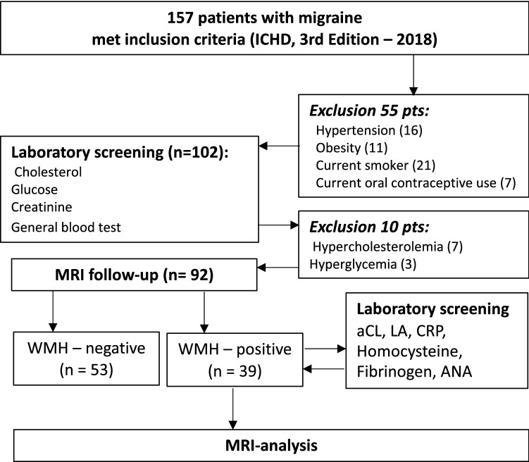
The diagnostic value of white matter hyperintensities (WMH) in different types of migraineare unknown. To evaluate the WMH pattern of different subtypes in migraine patients with no vascular risk factors. 92 migraine patients (73 females, mean age 34.6 ± 8.9; 61 episodic migraine, 31 chronic migraine, 36 migraine with aura, 56 migraine without aura) without vascular risk factors underwent brain MRI (3 T). We also included a matched healthy control group with no migraine (n = 24). The prevalence of WMH in different types of migraine was similar and ranged from 38.7 to 44.4%; the control group showed no WMH at all. Lesions were located within frontal, parietal and temporal lobes (in order of decreasing incidence) in juxtacortical and/or deep white matter. WMH appeared as round or slightly elongated foci with a median size of 2.5 mm [1.5; 3]. Total number, size and prevalence of WMH by lobes and white matter regions were similar between groups, and no interaction with age or sex was found. The number of lesions within the frontal lobe juxtacortical white matter correlated with the age of patients (r = 0.331, p = 0.001) and the duration since migraine onset (r = 0.264, p = 0.012). Patients with different migraine subtypes and without vascular risk factors are characterized by a similar pattern of WMH in the absence of subclinical infarctions or microbleedings. Therefore, WMH have no relevant prognostic value regarding the course of migraine and vascular complications. WMH pattern may be used to differentiate migraine as a primary disorder and other disorders with migraine-like headache and WMH.
Conflict of interest statement
The authors declare no competing interests.
Figures


References
-
- Global Burden of Disease Study Collaborators Global, regional, and national incidence, prevalence, and years lived with disability for 301 acute and chronic diseases and injuries in 188 countries, 1990–2013: A systematic analysis for the Global Burden of Disease Study 2013. Lancet. 2015;386:743–800. doi: 10.1016/S0140-6736(15)60692-4. - DOI - PMC - PubMed
-
- Headache Classification Committee of the International Headache Society (IHS) The International Classification of Headache Disorders. Cephalalgia. 2018;38:1–211. - PubMed
MeSH terms
Substances
LinkOut - more resources
Full Text Sources
Other Literature Sources
Medical
Miscellaneous
