. 2021 May 25;11(1):10837.
doi: 10.1038/s41598-021-90366-5.

Autopsy-diagnosed neurodegenerative dementia cases support the use of cerebrospinal fluid protein biomarkers in the diagnostic work-up

Affiliations

Autopsy-diagnosed neurodegenerative dementia cases support the use of cerebrospinal fluid protein biomarkers in the diagnostic work-up

Magdalena Bruzova et al. Sci Rep. .

Abstract

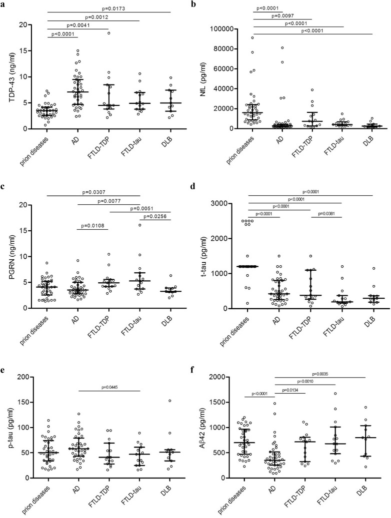
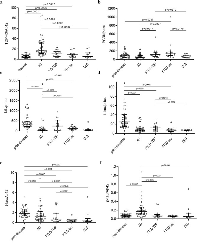
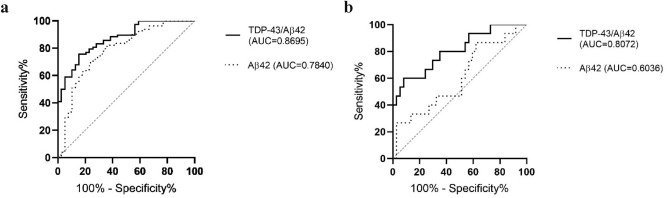
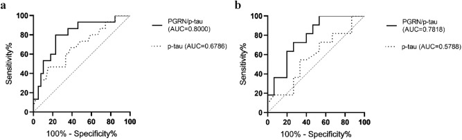
Various proteins play a decisive role in the pathology of different neurodegenerative diseases. Nonetheless, most of these proteins can only be detected during a neuropathological assessment, although some non-specific biomarkers are routinely tested for in the cerebrospinal fluid (CSF) as a part of the differential diagnosis of dementia. In antemortem CSF samples from 117 patients with different types of neuropathologically confirmed neurodegenerative disease with dementia, we assessed total-tau (t-tau), phosphorylated-tau (181P) (p-tau), amyloid-beta (1-42) (Aβ42), TAR DNA binding protein (TDP)-43, progranulin (PGRN), and neurofilament light (NfL) chain levels, and positivity of protein 14-3-3. We found t-tau levels and the t-tau/p-tau ratios were significantly higher in prion diseases compared to the other neurodegenerative diseases. Statistically significant differences in the t-tau/Aβ42 ratio predominantly corresponded to t-tau levels in prion diseases and Aβ42 levels in AD. TDP-43 levels were significantly lower in prion diseases. Additionally, the TDP-43/Aβ42 ratio was better able to distinguish Alzheimer's disease from other neurodegenerative diseases compared to using Aβ42 alone. In frontotemporal lobar degeneration, PRGN levels were significantly higher in comparison to other neurodegenerative diseases. There is an increasing need for biomarkers suitable for diagnostic workups for neurodegenerative diseases. It appears that adding TDP-43 and PGRN to the testing panel for neurodegenerative diseases could improve the resolution of differential diagnoses.


Conflict of interest statement

The authors declare no competing interests.

Figures

Figure 1
Figure 1
Scatter dot plots represent values of (a) TDP-43, (b) NfL, (c) PGRN, (d) t-tau, (e) p-tau, and (f) Aβ42 individually for prion diseases, AD, FTLD-TDP, FTLD-tau, and DLB. Bars represent medians with the interquartile range. Statistically significant p-values represent the exact two-tailed p-values of the Mann–Whitney test (p < 0.05). TDP-43, TAR DNA binding protein-43; PGRN, progranulin; NfL, neurofilament light chain; t-tau, total-tau; p-tau, phosphorylated-tau (181P); Aβ42, amyloid-beta (1–42); AD, Alzheimer disease; FTLD-TDP, frontotemporal lobar degeneration with TDP-43 inclusions; FTLD-tau, frontotemporal lobar degeneration with tau inclusions; DLB, dementia with Lewy bodies.
Figure 2
Figure 2
Scatter dot plots represent values of (a) TDP-43/Aβ42 ratio, (b) PGRN/p-tau ratio, (c) NfL/p-tau ratio, (d) t-tau/p-tau ratio, (e) t-tau/Aβ42 ratio, and (f) p-tau/Aβ42 ratio individually for prion diseases, AD, FTLD-TDP, FTLD-tau, and DLB. Bars represent medians with the interquartile range. Statistically significant p-values represent the exact two-tailed p-values of the Mann–Whitney test (p < 0.05). TDP-43, TAR DNA binding protein-43; NfL– neurofilament light chain; t-tau, total-tau; p-tau, phosphorylated-tau (181P); Aβ42, amyloid-beta (1–42); AD, Alzheimer disease; FTLD-TDP, frontotemporal lobar degeneration with TDP-43 inclusions; FTLD-tau, frontotemporal lobar degeneration with tau inclusions; DLB, dementia with Lewy bodies.
Figure 3
Figure 3
ROC diagrams of the TDP-43/Aβ42 ratio (solid line) and Aβ42 values (dotted line) in (a) AD vs. non-AD disease cases: for the TDP-43/Aβ42 ratio AUC 0.8695, CI 0.806 to 0.933, cut-off 6.737, p < 0.0001, for Aβ42 AUC 0.7840, CI 0.692 to 0.876, cut-off 425.5 pg/ml, p < 0.0001, (b) prion diseases vs. FTLD-TDP: for the TDP-43/Aβ42 ratio AUC 0.8072, CI 0.671 to 0.943, cut-off 9.537, p = 0.0006, for Aβ42 AUC 0.6036, CI 0.430 to 0.777, cut-off 867.0 pg/ml, p non-significant. TDP-43, TAR DNA binding protein-43; Aβ42, amyloid-beta (1–42); AD, Alzheimer disease; FTLD-TDP, frontotemporal lobar degeneration with TDP-43 inclusions; AUC, area under the curve; CI, 95% confidence interval.
Figure 4
Figure 4
ROC diagrams of the PGRN/p-tau ratio (solid line) and p-tau values (dotted line) in (a) AD vs. FTLD-tau: for the PGRN/p-tau ratio AUC 0.8000, CI 0.668 to 0.933, cut-off 92.1, p = 0.0007, for p-tau AUC 0.6786, CI 0.517 to 0.841, cut-off 36.0 pg/ml, p = 0.0436, (b) FTLD-tau vs. DLB: for the PGRN/p-tau ratio AUC 0.7818, CI 0.606 to 0.958, cut-off 137.6, p = 0.0158, for p-tau AUC 0.5788, CI 0.352 to 0.805, cut-off 50.5 pg/ml, p non-significant. PGRN, progranulin; p-tau, phosphorylated-tau (181P); AD, Alzheimer disease; FTLD-tau, frontotemporal lobar degeneration with tau inclusions; DLB, dementia with Lewy bodies; AUC, area under the curve; CI, 95% confidence interval.
Figure 5
Figure 5
ROC diagrams of protein 14–3-3 positivity, CI 0.753 to 0.934 (solid line), t-tau values higher than 1200 pg/ml, CI 0.868 to 0.984 (dashed line), and the combination of protein 14–3-3 positivity and t-tau values higher than 1200 pg/ml, CI 0.861 to 0.983 (dotted line) in prion vs. non- prion disease cases. t-tau, total tau; AUC, area under the curve; CI, 95% confidence interval.

References

    1. Golde TE, Miller VM. Proteinopathy-induced neuronal senescence: a hypothesis for brain failure in Alzheimer’s and other neurodegenerative diseases. Alzheimers Res. Ther. 2009;1:5. doi: 10.1186/alzrt5. - DOI - PMC - PubMed
    1. Golde TE, Borchelt DR, Giasson BI, Lewis J. Thinking laterally about neurodegenerative proteinopathies. J. Clin. Invest. 2013;123:1847–1855. doi: 10.1172/JCI66029. - DOI - PMC - PubMed
    1. Forman MS, Trojanowski JQ, Lee VM-Y. Neurodegenerative diseases: a decade of discoveries paves the way for therapeutic breakthroughs. Nat. Med. 2004;10:1055–1106. doi: 10.1038/nm1113. - DOI - PubMed
    1. Ross CA, Poirier MA. Protein aggregation and neurodegenerative disease. Nat. Med. 2004;10(Suppl):S10–S17. doi: 10.1038/nm1066. - DOI - PubMed
    1. Budka H, et al. Neuropathological diagnostic criteria for Creutzfeldt-Jakob disease (CJD) and other human spongiform encephalopathies (prion diseases) Brain Pathol. 1995;5:459–466. doi: 10.1111/j.1750-3639.1995.tb00625.x. - DOI - PubMed

Publication types

MeSH terms