Comparative Study
. 2021 May 25;11(1):10849.
doi: 10.1038/s41598-021-90380-7.

Gut microbiota composition in health-care facility-and community-onset diarrheic patients with Clostridioides difficile infection

Affiliations
Comparative Study

Gut microbiota composition in health-care facility-and community-onset diarrheic patients with Clostridioides difficile infection

Giovanny Herrera et al. Sci Rep. .

Abstract

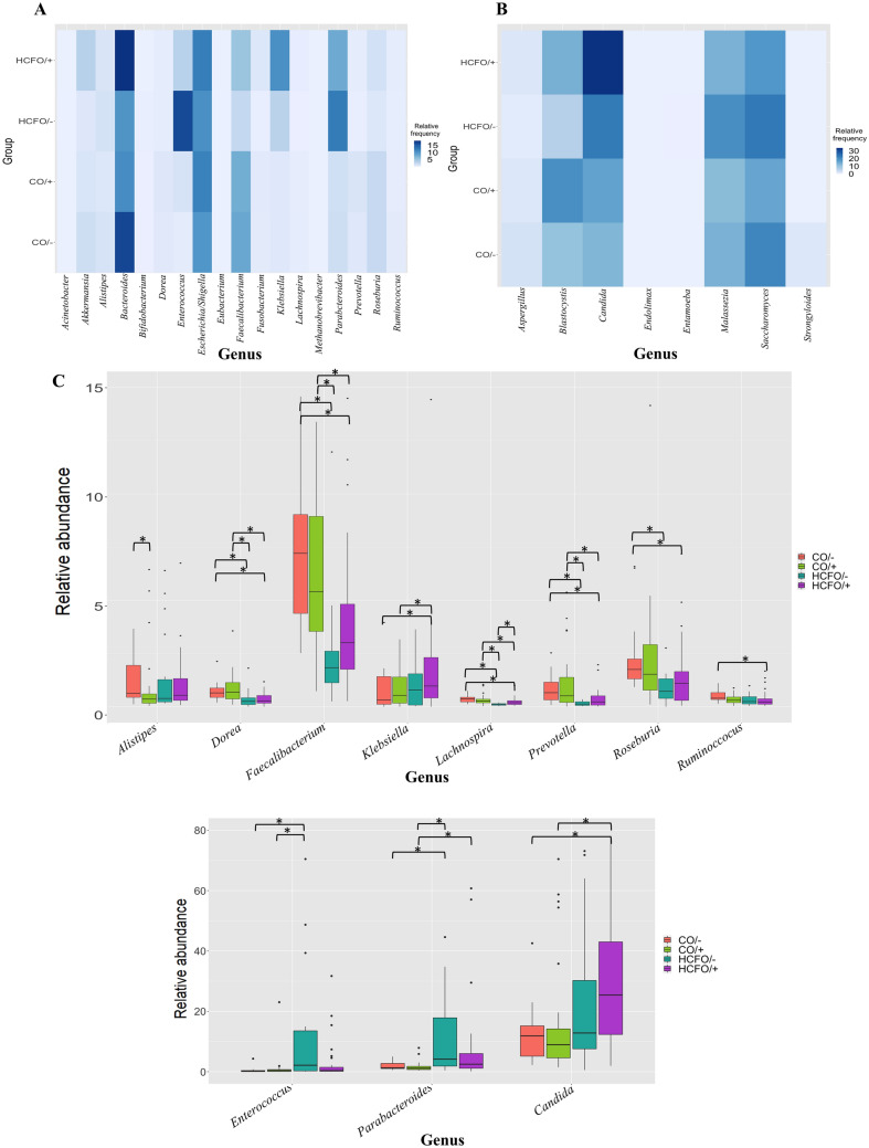
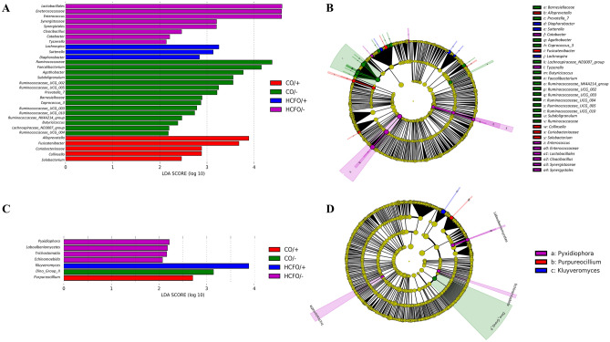
The role of gut microbiota in the establishment and development of Clostridioides difficile infection (CDI) has been widely discussed. Studies showed the impact of CDI on bacterial communities and the importance of some genera and species in recovering from and preventing infection. However, most studies have overlooked important components of the intestinal ecosystem, such as eukaryotes and archaea. We investigated the bacterial, archaea, and eukaryotic intestinal microbiota of patients with health-care-facility- or community-onset (HCFO and CO, respectively) diarrhea who were positive or negative for CDI. The CDI-positive groups (CO/+, HCFO/+) showed an increase in microorganisms belonging to Bacteroidetes, Firmicutes, Proteobacteria, Ascomycota, and Opalinata compared with the CDI-negative groups (CO/-, HCFO/-). Patients with intrahospital-acquired diarrhea (HCFO/+, HCFO/-) showed a marked decrease in bacteria beneficial to the intestine, and there was evidence of increased Archaea and Candida and Malassezia species compared with the CO groups (CO/+, CO/-). Characteristic microbiota biomarkers were established for each group. Finally, correlations between bacteria and eukaryotes indicated interactions among the different kingdoms making up the intestinal ecosystem. We showed the impact of CDI on microbiota and how it varies with where the infection is acquired, being intrahospital-acquired diarrhea one of the most influential factors in the modulation of bacterial, archaea, and eukaryotic populations. We also highlight interactions between the different kingdoms of the intestinal ecosystem, which need to be evaluated to improve our understanding of CDI pathophysiology.


Conflict of interest statement

The authors declare no competing interests.

Figures

Figure 1
Figure 1
Microbial composition of diarrheic patients’ gut microbiota by CDI status and by group (HCFO/+ , HCFO/−, CO/+, CO/−). (A) Bar plots showing the 9 major bacterial phyla by CDI status. (B) Bar plots of major eukaryotic groups by CDI status. (C) Bar plots of major bacterial phyla by group. (D) Bar plots of major eukaryotes by group. (E) Distribution of each bacterial phyla by CDI status. (F) Distribution of each bacterial phyla by group. (G) Distribution of eukaryotes by CDI status. (H) Distribution of eukaryotes by group. Figure created on R studio with ggplot package,.
Figure 2
Figure 2
Changes in bacteria and eukaryotes commonly found in the gut microbiota. (A) Heatmap of bacterial genera by group. (B) Heatmap of eukaryotic genera by group. (C) Boxplot showing the differences between groups by relative abundances of each genus. Statistical differences (Kruskall–Wallis test; Post-hoc: Dunn test with Benjamini–Hochberg correction and a confidence level of 95%) (p < 0.05) are indicated by an asterisk mark (*). Figure created on R studio with ggplot package,.
Figure 3
Figure 3
Possible interactions between kingdoms. Correlogram plots between bacteria and eukaryotes. ASVs corresponding to the most abundant phyla (Bacteroidetes, Firmicutes, Proteobacteria, Ascomycota and Basidiomycota) were compared. Outliers were deleted to only compare the ASVs corresponding to the most abundant genera (Spearman’s rho correlation method with Benjamini–Hochberg correction). Only were considered strong correlations (− 0.7 < ρ > 0.7; p value < 0.05). (A) CO/− group. (B) CO/+ group. (C) HCFO/−group. (D) HCFO/+ group. Figure created on R studio with psych package,.
Figure 4
Figure 4
Effect of size measurements (LEfSe) at phylum and genus levels. Linear discriminant analysis (LDA) combined with LEfSe showing a list of possible biomarkers that enable discrimination between groups. LDA ≥ 2.0 and p < 0.05 were considered significant in Kruskal–Wallis and pairwise Wilcoxon tests. (A) Distinct bacterial phyla and genera biomarkers by group. (B) Cladogram reporting the bacterial taxa (highlighted by small circles and shading) showing different abundance values in the groups. Circles’ diameters are proportional to the taxon’s abundance, and the shadow size is proportional to the effect size. (C) Distinct eukaryotic phyla and genera biomarkers by group. (D) Cladogram reporting eukaryotic taxa showing different abundance values in the groups. Figure created on Galaxy platform,.

Similar articles

Cited by

References

    1. Allegranzi B, et al. Burden of endemic health-care-associated infection in developing countries: Systematic review and meta-analysis. Lancet. 2011;377:228–241. doi: 10.1016/S0140-6736(10)61458-4. - DOI - PubMed
    1. Napolitano LM, Edmiston CE., Jr Clostridium difficile disease: Diagnosis, pathogenesis, and treatment update. Surgery. 2017;162:325–348. doi: 10.1016/j.surg.2017.01.018. - DOI - PubMed
    1. Zhu D, Sorg JA, Sun X. Clostridioides difficile biology: Sporulation, germination, and corresponding therapies for C. difficile infection. Front. Cell Infect. Microbiol. 2018;8:29. doi: 10.3389/fcimb.2018.00029. - DOI - PMC - PubMed
    1. Guh AY, Kutty PK. Clostridioides difficile infection. Ann. Intern. Med. 2018;169:ITC49–ITC64. doi: 10.7326/AITC201810020. - DOI - PMC - PubMed
    1. Dapa T, Unnikrishnan M. Biofilm formation by Clostridium difficile. Gut Microbes. 2013;4:397–402. doi: 10.4161/gmic.25862. - DOI - PMC - PubMed

Publication types

MeSH terms