. 2022 Mar;43(3):692-702.
doi: 10.1038/s41401-021-00691-8. Epub 2021 May 25.

Upregulation of wild-type p53 by small molecule-induced elevation of NQO1 in non-small cell lung cancer cells

Affiliations

Upregulation of wild-type p53 by small molecule-induced elevation of NQO1 in non-small cell lung cancer cells

Hong Yu et al. Acta Pharmacol Sin. 2022 Mar.

Abstract

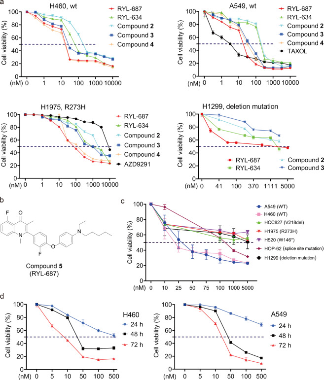
The tumor suppressor p53 is usually inactivated by somatic mutations in malignant neoplasms, and its reactivation represents an attractive therapeutic strategy for cancers. Here, we reported that a new quinolone compound RYL-687 significantly inhibited non-small cell lung cancer (NSCLC) cells which express wild type (wt) p53, in contract to its much weaker cytotoxicity on cells with mutant p53. RYL-687 upregulated p53 in cells with wt but not mutant p53, and ectopic expression of wt p53 significantly enhanced the anti-NSCLC activity of this compound. RYL-687 induced production of reactive oxygen species (ROS) and upregulation of Nrf2, leading to an elevation of the NAD(P)H:quinoneoxidoreductase-1 (NQO1) that can protect p53 by inhibiting its degradation by 20S proteasome. RYL-687 bound NQO1, facilitating the physical interaction between NQO1 and p53. NQO1 was required for RYL-687-induced p53 accumulation, because silencing of NQO1 by specific siRNA or an NQO1 inhibitor uridine, drastically suppressed RYL-687-induced p53 upregulation. Moreover, a RYL-687-related prodrug significantly inhibited tumor growth in NOD-SCID mice inoculated with NSCLC cells and in a wt p53-NSCLC patient-derived xenograft mouse model. These data indicate that targeting NQO1 is a rational strategy to reactivate p53, and RYL-687 as a p53 stabilizer bears therapeutic potentials in NSCLCs with wt p53.

Keywords: NQO1; RYL-687; non-small cell lung cancer; p53.


Conflict of interest statement

The authors declare no competing interests.

Figures

Fig. 1
Fig. 1. RYL-687 significantly inhibits lung cancer cells with wt p53.
a NSCLC cells were treated with different concentrations of indicated compounds for 72 h. Cell viability was evaluated by MTS assay. See Supplementary Table 1 for the structure of the compounds. b Chemical structures of RYL-687. c Seven lung cancer cell lines were treated with different concentrations of RYL-687 for 48 h, and cell viability was evaluated by MTS assay and is shown as relative viability compared with the control. d H460 and A549 cells were treated with the indicated concentrations of RYL-687 for the indicated time points, and cell viability was determined by MTS assay.
Fig. 2
Fig. 2. RYL-687 induces the expression of p53 in NSCLC cells.
a A549, H460, H1975 and H1299 cells were treated with the indicated concentrations of RYL-687 for 6 h, harvested, and subjected to Western blot analysis using the indicated antibodies. b After H460 cells were treated with RYL-687 for 6 h, nuclear and cytoplasmic protein fractions were prepared and evaluated by Western blotting. c Immunofluorescence assays of H460 cells using antibodies against p53 (green) and 4’,6-diamidino-2-phenylindole (DAPI) to counterstain the nucleus (blue) after treatment with RYL-687 at the indicated concentrations for the indicated time points. H460 (d) and A549 (e) cells were treated with RYL-687 (10 nM), Nutlin3A (10 μM), and cisplatin (DDP) (10 μM) at the indicated time points and lysed for Western blot analysis. f H460 cells were pre-treated with RYL-687 at 10 nM for 6 h to increase p53 to a detectable level, and then further treated with CHX (50 μg/mL) in the absence or presence of RYL-687 (10 nM), and the expression of p53 expression was evaluated by Western blotting. Numbers under the Western blot bands are the relative expression values to Actin determined by densitometry analysis. g H1299 cells were transfected with wt p53, treated with RYL-687, and analysed for cell viability. h H1975 cells were transfected with CRISPR-Cas9 system to silence the mutant p53, puromycin resistance cells were selected and transfected with wt p53, followed by treatment with RYL-687 and analysis of cell viability. **P < 0.01; ***P < 0.001; ****P < 0.0001.
Fig. 3
Fig. 3. Effects of RYL-687 on p53 target genes and cell fate.
a The cells were treated with RYL-687 at the indicated concentrations for 12 h, lysed, and subjected to immunoblotting using the indicated antibodies. b The mRNA levels of TP53, MDM2 and Bax were analyzed by quantitative RT-PCR, in H460 and A549 cells that were treated with RYL-687 at 10 nM for 12 h. c, d H460 and A549 cells were treated with the indicated concentrations of RYL-687 for 24 h. The cells were lysed for Western blot analysis using the indicated antibodies (c), or analysed by flow cytometry to evaluate the cell cycle distribution (d). e, f The cells were treated with the indicated concentrations of RYL-687 for 24 h, and lysed for Western blot analysis using total or cytosolic/mitochondrial proteins (e), or analysed by flow cytometry analysis using Annexin V-FITC/PI staining for apoptotic cell death (f). g Colony formation assays were performed in H460 and A549 cells with the indicated concentrations of RYL-687. Representative images are shown. *P < 0.05, **P < 0.01; ***P < 0.001 and ****P < 0.0001.
Fig. 4
Fig. 4. RYL-687 promotes NQO1-p53 interaction and reactive oxygen species production.
a Silver staining of proteins precipitated with Biotin and Bio-687 at 10 μM for 6 h in H460 cells. b H460 cells were transfected with or without siNQO1, treated with Biotin or Bio-687 at 10 μM for 6 h and lysed, and the cell lysates were subjected to immunoprecipitation using streptavidin agarose and Western blotting using indicated antibodies. c H460 cells were treated with RYL-687 at 10 nM for 6 h, lysed, and subjected to immunoprecipitation and immunoblotting using indicated antibodies. d The expression of p53 and NQO1 in H460 and A549 cells treated with vehicle control (DMSO) or RYL-687 at 10–50 nM for 12 h. The cells were harvested and detected by immunofluorescence assays with indicated antibodies. e H460 cells were treated with RYL-687 at 10 nM for 6–12 h and lysed, RNA was extracted, and NQO1 expression was evaluated by quantitative RT-PCR. P values, Student’s t test, *P < 0.05; **P < 0.01, ***P < 0.001. f Western blot analysis of NQO1, Nrf2 and p53 expression in H460 and A549 cells treated with RYL-687 for 12 h. g H460 and A549 cells were incubated with RYL-687 for 24 h. ROS detection was performed with DCFH-DA dye in different groups by flow cytometry. h H460 and A549 cells were treated with the indicated concentrations of RYL-687 for 12 h, lysed, and analysed by Western blot using indicated antibodies.
Fig. 5
Fig. 5. NQO1 is required for RYL-687-induced upregulation of p53.
a H460 cells were exposed to RYL-687 and RYL-687 in combination with uridine (50 μM) for 48 h. Cell viability was evaluated by MTS assay. b H460 and A549 cells were treated with RYL-687 and RYL-687 plus uridine for 24 h. The cells were analysed by flow cytometry to evaluate the cell cycle distribution. c H460 cells were transfected with siNQO1, treated with RYL-687 at 10 nM for 12 h, and lysed for Western blot analysis using indicated antibodies. d Western blot analysis of p53 levels in H460 and A549 cells treated with RYL-687 and/or uridine (50 μM) for 12 h.
Fig. 6
Fig. 6. The prodrug exhibits potent anti-lung cancer activity in vivo.
a H460 (1 × 106) cells were inoculated subcutaneously into the right flank of the NOD-SCID mice, which were treated with indicated concentrations of prodrug and MTX. Tumor volume was estimated every 2 days. Data are shown as mean ± SD. b Images of xenograft tumors isolated from the mice. c Weights of xenograft tumors isolated from the mice. d Body weights of the mice. e Patient-derived xenograft mouse model was established using NOD-SCID mice and a wt-p53-bearing NSCLC tumor sample and treated with prodrug and MTX, and tumor volume was estimated every 2 days. Data are shown as mean ± SD. n = 5 for each group. f Images of xenograft tumors obtained from the mice. g Weights of xenograft tumors isolated from the mice. h Body weights of the mice. CDX cell-derived xenograft, PDX patient-derived xenograft. *P < 0.05; **P < 0.01.
Fig. 7
Fig. 7. RYL-687 is a p53 re-activator.
Schematic representation of the mechanism of the action of RYL-687 in NSCLC cells with wt p53.

Similar articles

Cited by

References

    1. Siegel RL, Miller KD, Fuchs HE, Jemal A. Cancer statistics, 2021. CA Cancer J Clin. 2021;71:7–33. doi: 10.3322/caac.21654. - DOI - PubMed
    1. Howlader N, Forjaz G, Mooradian MJ, Meza R, Kong CY, Cronin KA, et al. The effect of advances in lung-cancer treatment on population mortality. N Engl J Med. 2020;383:640–9. - PMC - PubMed
    1. Herbst RS, Morgensztern D, Boshoff C. The biology and management of non-small cell lung cancer. Nature. 2018;553:446–54. - PubMed
    1. Hirsch FR, Scagliotti GV, Mulshine JL, Kwon R, Curran WJ, Jr., Wu YL, et al. Lung cancer: current therapies and new targeted treatments. Lancet. 2017;389:299–311. - PubMed
    1. Siegel RL, Miller KD, Jemal A. Cancer statistics, 2019. CA Cancer J Clin. 2019;69:7–34. - PubMed

MeSH terms