. 2021 May 4:2021:5562340.
doi: 10.1155/2021/5562340. eCollection 2021.

Assessment of the Vanillin Anti-Inflammatory and Regenerative Potentials in Inflamed Primary Human Gingival Fibroblast

Affiliations

Assessment of the Vanillin Anti-Inflammatory and Regenerative Potentials in Inflamed Primary Human Gingival Fibroblast

Erica Costantini et al. Mediators Inflamm. .

Abstract

Background: Inflammatory responses have been associated with delayed oral mucosal wound healing and the pathogenesis of the periodontal disease. The invasion of microbes into the tissues and the establishment of a chronic infection may be due to impaired healing. The protracted inflammatory phase may delay wound healing and probably support tissue fibrosis and reduce tissue regeneration. Vanillin is a well-known natural compound with potential anti-inflammatory capacity. Hence, we hypothesized that Vanillin could accelerate wound healing reducing inflammation and especially cytokine production making the oral tissue repair process easier.

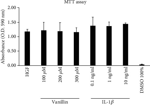
Methods: Our hypothesis was tested using primary human gingival fibroblast (HGF) cell pretreated with Vanillin and primed with IL-1β, as inductor of proinflammatory environment. After 24 hours of treatments, the gene expression and production of IL-6, TNF-α, IL-8, COX-2, iNOS, and nitric oxide (NO) generation and the wound healing rate were determined.

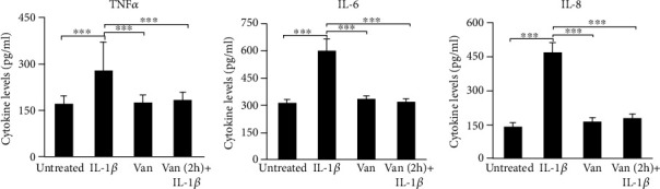
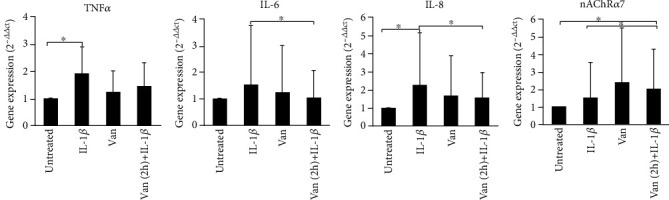
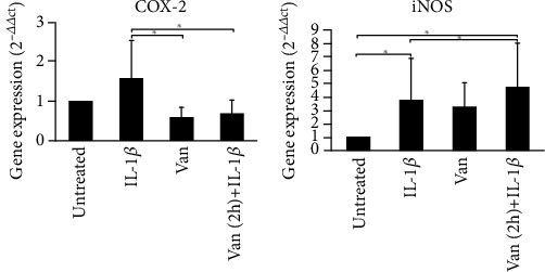
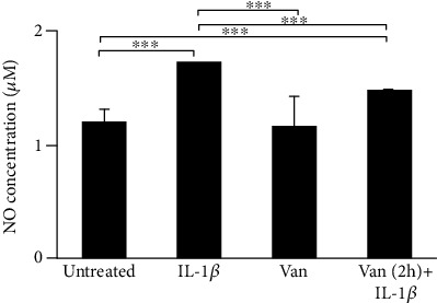
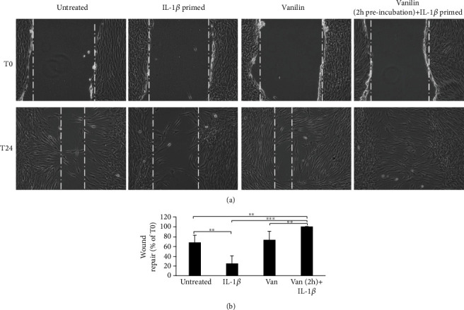
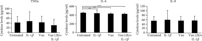
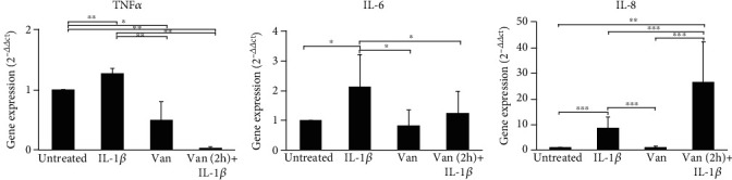
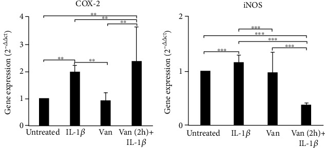
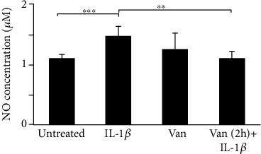
Results: In IL-1β-primed cells, preincubation with Vanillin reduced IL-6, IL-8, COX-2, and iNOS expression and NO release, compared to IL-1β-primed cells. Moreover, Vanillin determines the increased gene expression of nAChRα7, leading us to hypothesize a role of Vanillin in the activation of the cholinergic anti-inflammatory pathway. Furthermore, in presence of mechanical injury, the Vanillin preincubation, wound closure may be reducing the expression and release of IL-6 and TNF-α and upregulation of COX-2 and IL-8.

Conclusion: Together, the results of this study highlight the anti-inflammatory and tissue repair ability of Vanillin in IL-1β-primed HGF. Therefore, Vanillin shows a potential therapeutic interest as an inflammatory modulator molecule with novel application in periodontal regeneration and oral health.


Conflict of interest statement

The authors declare no conflict of interest.

Figures

Figure 1
Figure 1
MTT viability assay in HGF cell treated for 24 h with Vanillin (100, 200, and 300 μM) or IL-1β (0.1, 1, and 10 ng/ml). Absorbance values are given as media ±SD of three independent experiments.
Figure 2
Figure 2
TNF-α, IL-6, and IL-8 levels in HGF cell culture supernatant. The mean values ± SD were reported, ∗∗∗p < 0.001. All experiments were repeated in triplicate.
Figure 3
Figure 3
Gene expression of TNF-α, IL-6, IL-8, and nAChRα7 in HGF cells. Data are reported as mean and 95% CI, of three independent experiments. p < 0.05. IL-1β: IL-1β-primed cells; Van: Vanillin; Van (2 h)+IL-1β: Vanillin (2 h pretreatment) plus IL-1β.
Figure 4
Figure 4
Gene expression of COX-2 and iNOS in HGF cells. Data are reported as mean and 95% CI, of three independent experiments, p < 0.05. IL-1β: IL-1β-primed cells; Van: Vanillin; Van (2 h)+IL-1β: Vanillin (2 h pre-treatment) plus IL-1β.
Figure 5
Figure 5
Levels of NO in HGF cells were determined based on Griess reagent reaction. Experiments were conducted in duplicate, and changes were reported as mean ± SD; ∗∗∗p < 0.001. IL-1β: IL-1β-primed cells; Van: Vanillin; Van (2 h)+IL-1β: Vanillin (2 h pretreatment) plus IL-1β.
Figure 6
Figure 6
Wound healing of scratched oral fibroblast monolayer photographs and measurement of wound area were made immediately after the scratch (T0) and after 24 h (T24). (a) Representative images of wound-healing assay. (b) Wound repair was evaluated measuring the remaining cell-free area after 24 h and expressed as a percentage of the initial wound size (T0) assumed as 100%. ∗∗p < 0.01; ∗∗∗p < 0.001. IL-1β: IL-1β-primed cells; Van: Vanillin; Van (2 h)+IL-1β: Vanillin (2 h pretreatment) plus IL-1β.
Figure 7
Figure 7
Levels of TNF-α, IL-6, and IL-8 in supernatant of HGF scratched cells. Data are reported as mean and 95% CI, of three independent experiments. ∗∗p < 0.01; ∗∗∗p < 0.001. IL-1β: IL-1β-primed cells; Van: Vanillin; Van (2 h)+IL-1β: Vanillin (2 h pretreatment) plus IL-1β.
Figure 8
Figure 8
Gene expression of TNF-α, IL-6, and IL-8 in HGF scratched cells. Data are reported as mean and 95% CI, of three independent experiments. p < 0.05; ∗∗p < 0.01; ∗∗∗p < 0.001. IL-1β: IL-1β-primed cells; Van: Vanillin; Van (2 h)+IL-1β: Vanillin (2 h pretreatment) plus IL-1β.
Figure 9
Figure 9
Gene expression of COX-2 and iNOS in HGF cells. Data are reported as mean and 95% CI, of three independent experiments. ∗∗p < 0.01; ∗∗∗p < 0.001. IL-1β: IL-1β-primed cells; Van: Vanillin; Van (2 h)+IL-1β: Vanillin (2 h pretreatment) plus IL-1β.
Figure 10
Figure 10
Levels of NO in HGF cells were determined based on Griess reagent reaction. Experiments were conducted in duplicate, and changes were reported as mean ± SD. ∗∗p < 0.01; ∗∗∗p < 0.001. IL-1β: IL-1β-primed cells; Van: Vanillin; Van (2 h)+IL-1β: Vanillin (2 h pretreatment) plus IL-1β.

References

    1. Chen L., Deng H., Cui H., et al. Inflammatory responses and inflammation-associated diseases in organs. Oncotarget. 2018;9(6, article 23208):7204–7218. doi: 10.18632/oncotarget.23208. - DOI - PMC - PubMed
    1. Kany S., Vollrath J. T., Relja B. Cytokines in inflammatory disease. International Journal of Molecular Sciences. 2019;20(23):p. 6008. doi: 10.3390/ijms20236008. - DOI - PMC - PubMed
    1. Reale M., Velluto L., di Nicola M., et al. Cholinergic markers and cytokines in OSA patients. International Journal of Molecular Sciences. 2020;21(9):p. 3264. doi: 10.3390/ijms21093264. - DOI - PMC - PubMed
    1. Pihlstrom B. L., Michalowicz B. S., Johnson N. W. Periodontal diseases. Lancet. 2005;366(9499):1809–1820. doi: 10.1016/S0140-6736(05)67728-8. - DOI - PubMed
    1. Palombo E. A. Traditional medicinal plant extracts and natural products with activity against oral bacteria: potential application in the prevention and treatment of oral diseases. Evidence-based complementary and Alternative Medicine. 2011;2011:15. doi: 10.1093/ecam/nep067.680354 - DOI - PMC - PubMed