Case Reports
. 2021 Apr 19;13(4):e14570.
doi: 10.7759/cureus.14570.

Atrophic Dermatofibroma: A Unique Dermatofibroma Variant

Affiliations
Case Reports

Atrophic Dermatofibroma: A Unique Dermatofibroma Variant

Nikolas Gutierrez et al. Cureus. .

Abstract

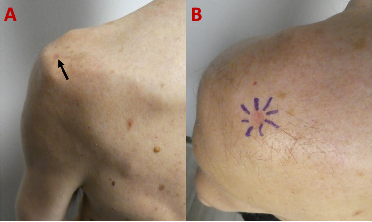
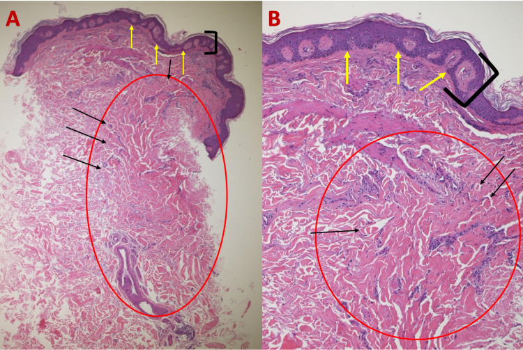
Dermatofibromas are benign skin tumors with several variants, including the rare, uncommonly described atrophic dermatofibroma. To the best of our knowledge, there are currently 105 reported cases of atrophic dermatofibromas in the literature. This variant typically presents as a flat or depressed macule whose color can range from brown to white to red; in contrast to classic dermatofibromas that typically occur on the legs, atrophic dermatofibromas have a tendency to occur on the upper back and arms. An atrophic dermatofibroma can be clinically diagnosed; however, given the broad spectrum of clinical features of this lesion, a biopsy may be required. Characteristic pathologic features include epidermal acanthosis, basilar hyperpigmentation, fibroblast hyperplasia, and decreased or absent elastic fibers within the lesion. The pathogenesis of this lesion is not yet fully understood; however, it has been postulated that the loss of elastic fibers plays a key role in its development and characteristic atrophic appearance. We present the cases of two men with biopsy-confirmed atrophic dermatofibromas: a 47-year-old man with a pigmented macule on the right upper back and a 68-year-old man with an erythematous patch on the left posterolateral shoulder. The clinical and pathologic features of atrophic dermatofibromas are also summarized.

Keywords: acanthosis; atrophic; atrophy; dermatofibroma; elastic; fibers; fibroblast; hyperpigmentation.


Conflict of interest statement

Philip R. Cohen is a consultant for ParaPRO.

Figures

Figure 1
Figure 1. Clinical presentation of an atrophic dermatofibroma.
Distant (A) and closer (B) views of a 47-year-old man with an atrophic dermatofibroma presenting as a flat, atrophic, hyperpigmented macule (black arrow) on his right upper back.
Figure 2
Figure 2. Microscopic presentation of H&E-stained sections of an atrophic dermatofibroma on the right upper back of a 47-year-old man.
Low (A) and higher (B) magnification of H&E-stained sections of an atrophic dermatofibroma shows a central depression (between blue arrows), epidermal acanthosis (thickening of the epidermis as shown between black bracket), basilar hyperpigmentation (yellow arrows), and a fibroblast proliferation (red circle) with trapped collagen bundles at the periphery (black arrows) (H&E: A, ×5; B, ×20). H&E, hematoxylin and eosin
Figure 3
Figure 3. Microscopic presentation of VVG-stained sections of an atrophic dermatofibroma on the right upper back of a 47-year-old man.
Low (A) and higher (B) magnification of VVG-stained sections of an atrophic dermatofibroma reveals a loss of black-stained elastic fibers (blue circle) within the lesion; in contrast, elastic fibers (black arrows) can be seen in the dermis above, adjacent, and deep to the dermal tumor. The red circle of image A is shown at higher magnification in image B (VVG: A, ×5; B, ×40). VVG, Verhoeff-van Gieson
Figure 4
Figure 4. Atrophic dermatofibroma on the left posterolateral shoulder of a 68-year-old man.
The distant (A) view of an atrophic dermatofibroma presenting as a depressed, erythematous patch (black arrow) on the left posterolateral shoulder. A closer view (B) demonstrates the peripheral edges of the central, atrophic tumor outlined by purple pen markings prior to biopsy specimen collection.
Figure 5
Figure 5. Histologic features of atrophic dermatofibroma on the left shoulder of a 68-year-old man on H&E sections.
Low (A) and higher (B) magnification of H&E-stained sections of an atrophic dermatofibroma showing epidermal acanthosis (thickening of the epidermis as shown between black bracket), basilar hyperpigmentation (yellow arrows), and proliferation of dermal fibroblasts (red circle) with trapped collagen bundles (black arrows) (H&E: A, ×5; B, ×20). H&E, hematoxylin and eosin
Figure 6
Figure 6. VVG-stained sections of an atrophic dermatofibroma on the left shoulder of a 68-year-old man.
Low (A) and higher (B) magnification of VVG-stained sections of an atrophic dermatofibroma reveals an absence of elastic fibers within the tumor (red circle); however, black-stained elastic fibers (yellow arrows) can be visualized in the deep dermis above, adjacent, and deep to the dermatofibroma (VVG: A, ×5; B, ×20). VVG, Verheoff-van Gieson

References

    1. Atrophic variants of dermatofibroma and dermatofibrosarcoma protuberans. Zelger BW, Ofner D, Zelger BG. Histopathology. 1995;26:519–527. - PubMed
    1. Atrophic dermatofibroma: a comprehensive literature review. Cohen PR, Erickson CP, Calame A. Dermatol Ther (Heidelb) 2019;9:449–468. - PMC - PubMed
    1. Atrophic dermatofibroma and dermatofibrosarcoma protuberans. Page EH, Assaad DM. J Am Acad Dermatol. 1987;17:947–950. - PubMed
    1. Atrophic dermatofibroma: a case report and review of the literature. Hendi A, Jukic DM, Kress DW, Brodland DG. Dermatol Surg. 2002;28:1085–1087. - PubMed
    1. A rare case of atrophic dermatofibroma with dermoscopic findings. Aktaş Karabay E, Demir D, Gürsoy F, Zindancı İ. J Cosmet Dermatol. 2021 - PubMed

Publication types

LinkOut - more resources