. 2021 Mar 21;3(3):100281.
doi: 10.1016/j.jhepr.2021.100281. eCollection 2021 Jun.

A human liver chimeric mouse model for non-alcoholic fatty liver disease

Affiliations

A human liver chimeric mouse model for non-alcoholic fatty liver disease

Beatrice Bissig-Choisat et al. JHEP Rep. .

Abstract

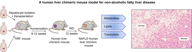
Background & aims: The accumulation of neutral lipids within hepatocytes underlies non-alcoholic fatty liver disease (NAFLD), which affects a quarter of the world's population and is associated with hepatitis, cirrhosis, and hepatocellular carcinoma. Despite insights gained from both human and animal studies, our understanding of NAFLD pathogenesis remains limited. To better study the molecular changes driving the condition we aimed to generate a humanised NAFLD mouse model.

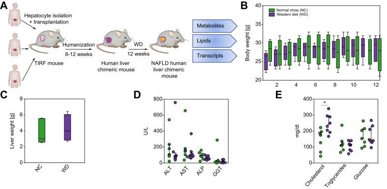
Methods: We generated TIRF (transgene-free Il2rg -/-/Rag2 -/-/Fah -/-) mice, populated their livers with human hepatocytes, and fed them a Western-type diet for 12 weeks.

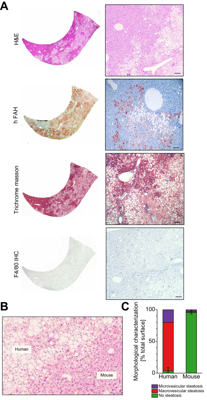
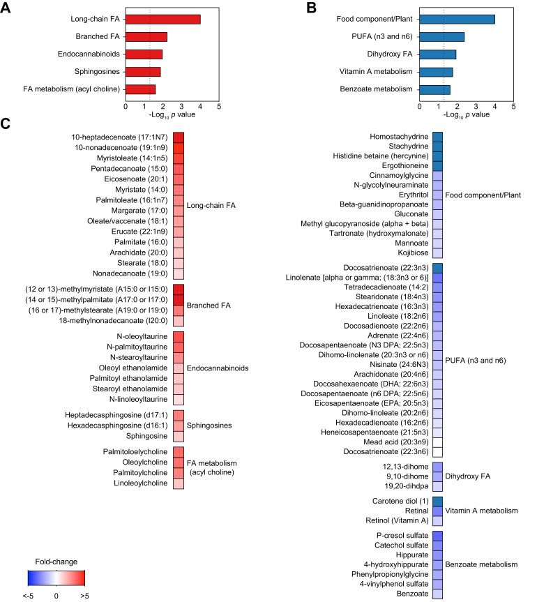
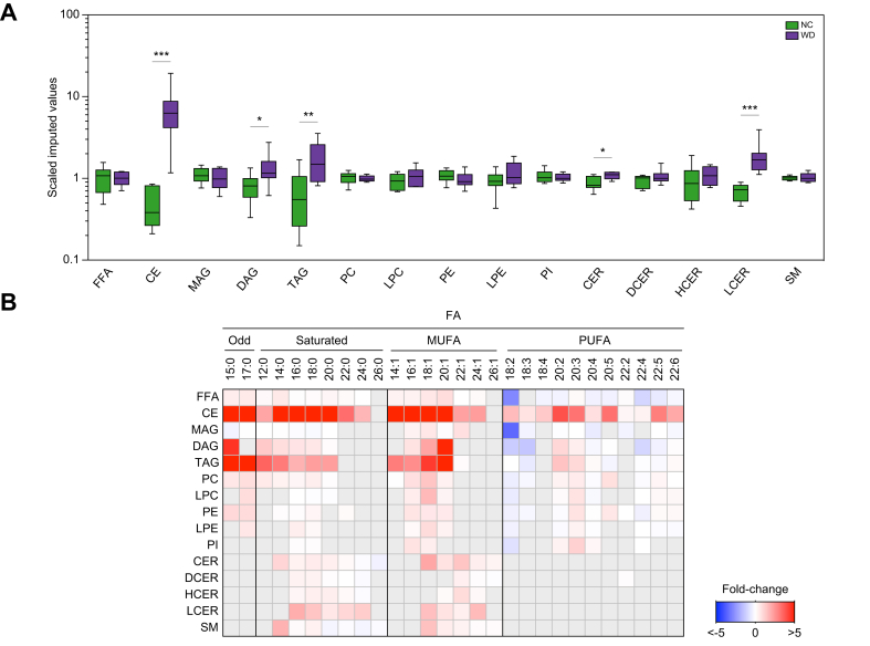
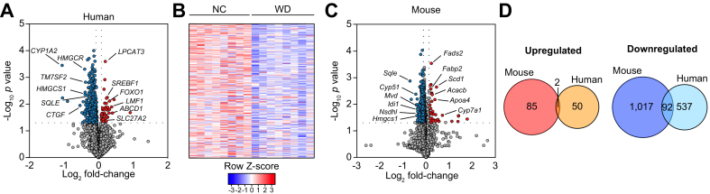
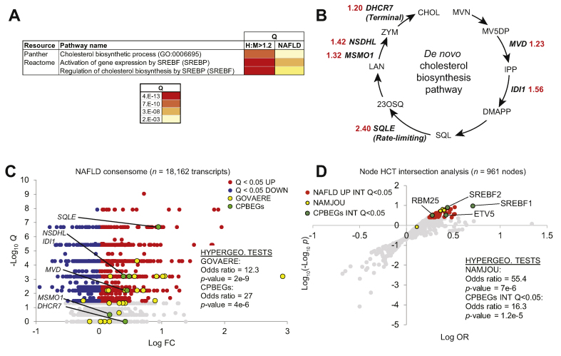
Results: Within the same chimeric liver, human hepatocytes developed pronounced steatosis whereas murine hepatocytes remained normal. Unbiased metabolomics and lipidomics revealed signatures of clinical NAFLD. Transcriptomic analyses showed that molecular responses diverged sharply between murine and human hepatocytes, demonstrating stark species differences in liver function. Regulatory network analysis indicated close agreement between our model and clinical NAFLD with respect to transcriptional control of cholesterol biosynthesis.

Conclusions: These NAFLD xenograft mice reveal an unexpected degree of evolutionary divergence in food metabolism and offer a physiologically relevant, experimentally tractable model for studying the pathogenic changes invoked by steatosis.

Lay summary: Fatty liver disease is an emerging health problem, and as there are no good experimental animal models, our understanding of the condition is poor. We here describe a novel humanised mouse system and compare it with clinical data. The results reveal that the human cells in the mouse liver develop fatty liver disease upon a Western-style fatty diet, whereas the mouse cells appear normal. The molecular signature (expression profiles) of the human cells are distinct from the mouse cells and metabolic analysis of the humanised livers mimic the ones observed in humans with fatty liver. This novel humanised mouse system can be used to study human fatty liver disease.

Keywords: ALP, alkaline phosphatase; ALT, alanine aminotransferase; AST, aspartate aminotransferase; CBPEGs, cholesterol biosynthesis pathway enzyme genes; CE, cholesteryl ester; CER, ceramide; CHHs, chimeric human hepatocytes; CMHs, chimeric mouse hepatocytes; CT, confidence transcript; DAG, diacylglycerol; DCER, dihydroceramide; DEG, differentially expressed gene; FA, fatty acid; FAH, fumarylacetoacetate hydrolase; FFA, free fatty acid; GGT, gamma-glutamyl transpeptidase; HCC, hepatocellular carcinoma; HCER, hexosylceramide; HCT, high confidence transcriptional target; Human disease modelling; Humanised mice; LCER, lactosylceramide; LPC, lysophosphatidylcholine; LPE, lysophosphatidylethanolamine; Lipid metabolism; MAG, monoacylglycerol; MUFA, monounsaturated fatty acid; NAFLD, non-alcoholic fatty liver disease; NASH, non-alcoholic steatohepatitis; NC, normal chow; NTBC, nitisinone; Non-alcoholic fatty liver disease; PC, phosphatidylcholine; PE, phosphatidylethanolamine; PI, phosphatidylinositol; PNPLA3, patatin-like-phospholipase domain-containing protein 3; PUFA, polyunsaturated free FA; SM, sphingomyelin; SREBP, sterol regulatory element-binding protein; Steatosis; TAG, triacylglycerol; TIRF, transgene-free Il2rg-/-/Rag2-/-/Fah-/-; WD, Western-type diet; hALB, human albumin.


Conflict of interest statement

The authors declare no personal or financial conflicts of interest. Please refer to the accompanying ICMJE disclosure forms for further details.

Figures

None
Graphical abstract
Fig 1
Fig 1
Experimental set-up and basic parameters of humanised TIRF mice on Western-type diet (WD). (A) TIRF mice were transplanted with 3 different samples of human hepatocytes. After reaching high human chimerism, animals were placed on WD or NC for 12 weeks, after which they were euthanised for transcriptomic, metabolomic, and lipidomic analyses of the human liver cells. (B) Body weights (n = 6–8 per group), (C) liver weights (n = 6–8 per group), and (D,E) blood chemistry (n = 6–8 per group) after 12 weeks. (E) Plasma concentrations of cholesterol, triacylglycerol, and glucose after 12 weeks of diet. Data are presented as mean ± SD. p <0.05 using Student's t-test. ALP, alkaline phosphatase; ALT, alanine aminotransferase; AST, aspartate aminotransferase; GGT, gamma-glutamyl transpeptidase; NAFLD, non-alcoholic fatty liver disease; NC, normal chow; TIRF, transgene-free Il2rg-/-/Rag2-/-/Fah-/-.
Fig 2
Fig 2
Diet-induced steatosis is associated with human hepatocytes in NAFLD xenograft mice. Histological analyses were performed in liver sections after 12 weeks of Western-type diet (n = 6–8 per group). (A) Representative images of H&E-, fumarylacetoacetate hydrolase (FAH)-, Trichrome Masson-stained and F4/80-immunostained liver sections. Scale bar = 50 μm. (B) Representative image of H&E-stained liver sections showing separation of hepatocytes from human or murine origin (dotted line). Scale bar = 50 μm. (C) Quantification of macro- and microvesicular steatosis in human and murine liver tissue. Percentage surface area of multiple lobes (2–5) of NAFLD xenograft mice (n = 6) are given with whiskers for the range of non-steatotic human or murine hepatocytes (see methods for details). NAFLD, non-alcoholic fatty liver disease.
Fig 3
Fig 3
NAFLD xenograft mouse livers show altered metabolic profile after 12 weeks on WD. Global metabolic profiles were determined in livers of 12 week WD or NC chimeric mice (n = 8 per group). (A) Metabolite species increased in livers of NAFLD xenograft mice after 12 weeks of WD. (B) Metabolite species decreased in livers of NAFLD xenograft mice after 12 weeks on WD. (C) Relative levels of individual metabolites are shown. FA, fatty acid; NAFLD, non-alcoholic fatty liver disease; NC, normal chow; PUFA, polyunsaturated fatty acid; WD, Western-type diet.
Fig 4
Fig 4
Altered lipid profile in livers of NAFLD xenograft mice. Global lipid profiles were determined in livers of 12-week WD or NC chimeric mice (n = 8 per group). (A) Hepatic levels of lipids. ∗p <0.05, ∗p <0.01, and ∗∗∗p <0.001 using Welch's t-test. (B) Hepatic fatty acyl composition (relative to NC-fed humanised mice). Grey boxes, data not available. CE, cholesteryl ester; CER, ceramide; DAG, diacylglycerol; DCER, dihydroceramide; FFA, free fatty acid; HCER, hexosylceramide; LCER, lactosylceramide; LPC, lysophosphatidylcholine; LPE, lysophosphatidylethanolamine; MAG, monoacylglycerol; NAFLD, non-alcoholic fatty liver disease; NC, normal chow; PC, phosphatidylcholine; PE, phosphatidylethanolamine; PI, phosphatidylinositol; TAG, triacylglycerol; WD, Western-type diet.
Fig 5
Fig 5
Divergent transcriptional responses to WD between CHHs and CMHs in NAFLD xenograft mice. Transcriptomic analysis of human (A and B) and mouse (C and D) hepatocytes was performed in livers of 12 week WD or NC chimeric mice (n = 8 per group). Upregulated genes in the WD-fed mice relative to the NC group are shown in red, downregulated genes are in blue. (D) Groupwise comparison of differentially expressed mouse and human genes (WD vs. NC). CHHs, chimeric human hepatocytes; CMHs, chimeric mouse hepatocytes; NC, normal chow; NAFLD, non-alcoholic fatty liver disease; WD, Western-type diet.
Fig 6
Fig 6
Distinct human and mouse cholesterol biosynthesis enzyme expression profiles in chimeric livers recapitulate clinical NAFLD. (A) Enrichment of SREBP1-cholesterol biosynthesis transcriptional pathways connects chimeric human hepatocytes (CHHs) and clinical NAFLD. (B) H:M >1.2 genes encode enzymes in the de novo cholesterol biosynthesis pathway. Enzyme names are shown in (D). Red numerals refer to CHH:CMH relative expression ratios. (C) The human NAFLD transcriptomic consensome ranks 18,162 genes based on their discovery rates across 20 independent, publicly archived clinical NAFLD case:control transcriptomic datasets. Hypergeometric test statistics for over-representation of the 25-gene NAFLD severity signature (GOVAERE) and CBPEGs among NAFLD CTs (mean case: control FC >1.25) are indicated. Q, FDR-corrected consensome p-value. (D) Node HCT intersection analysis of CPBEGs and FC >1.2 Q <0.05 clinical NAFLD consensome genes. NAFLD UP INT Q<0.05: nodes with significant (Q <0.05) intersections with NAFLD consensome CTs with mean case:control FC>1.2. CPBEGs INT Q <0.05: nodes with significant (Q <0.05) intersections with CPBEGs. Full data are in Table S6, section 4. CBPEG, cholesterol biosynthesis pathway enzyme genes; FC, fold change; FDR, false discovery rate; HCT, high confidence transcriptional target; NAFLD, non-alcoholic fatty liver disease; OR, odds ratio; SREBP1, sterol regulatory element-binding protein 1.

Similar articles

Cited by

References

    1. Younossi Z.M., Koenig A.B., Abdelatif D., Fazel Y., Henry L., Wymer M. Global epidemiology of nonalcoholic fatty liver disease – meta-analytic assessment of prevalence, incidence, and outcomes. Hepatology. 2016;64:73–84. - PubMed
    1. Alves-Bezerra M., Cohen D.E. Triglyceride metabolism in the liver. Compr Physiol. 2017;8:1–8. - PMC - PubMed
    1. Bessone F., Razori M.V., Roma M.G. Molecular pathways of nonalcoholic fatty liver disease development and progression. Cell Mol Life Sci. 2019;76:99–128. - PMC - PubMed
    1. Font-Burgada J., Sun B., Karin M. Obesity and cancer: the oil that feeds the flame. Cell Metab. 2016;23:48–62. - PubMed
    1. Hebbard L., George J. Animal models of nonalcoholic fatty liver disease. Nat Rev Gastroenterol Hepatol. 2011;8:35–44. - PubMed