Review
. 2021 Feb 25:11:100101.
doi: 10.1016/j.mtbio.2021.100101. eCollection 2021 Jun.

Recent developments in bio-scaffold materials as delivery strategies for therapeutics for endometrium regeneration

Affiliations
Review

Recent developments in bio-scaffold materials as delivery strategies for therapeutics for endometrium regeneration

X Li et al. Mater Today Bio. .

Abstract

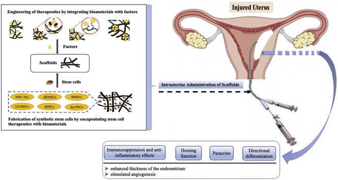
Intrauterine adhesions (IUAs) refer to the repair disorder after endometrial injury and may lead to uterine infertility, recurrent miscarriage, abnormal menstrual bleeding, and other obstetric complications. It is a pressing public health issue among women of childbearing age. Presently, there are limited clinical treatments for IUA, and there is no sufficient evidence that these treatment modalities can effectively promote regeneration after severe endometrial injury or improve pregnancy outcome. The inhibitory pathological micro-environment is the main factor hindering the repair of endometrial damaged tissues. To address this, tissue engineering and regenerative medicine have been achieving promising developments. Particularly, biomaterials have been used to load stem cells or therapeutic factors or construct an in situ delivery system as a treatment strategy for endometrial injury repair. This article comprehensively discusses the characteristics of various bio-scaffold materials and their application as stem cell or therapeutic factor delivery systems constructed for uterine tissue regeneration.

Keywords: Asherman's syndrome/endometrium regeneration; BMNCs, autologous bone marrow mononuclear cells; BMSCs, bone marrow mesenchymal stem cells; Biological scaffold material; D&C, Dilatation and curettage; ECM, extracellular matrix; En-PSC, endometrial perivascular cells; IUA, Intrauterine adhesions; KGF, Keratinocyte growth factor; MSC-Sec, Mesenchymal stem cell-secretome; SDF-1α, stromal cell-derived factor-1α; Scaffold-based therapeutics delivery systems; Stem cell; Therapeutic factor; UCMSCs, umbilical cord derived mesenchymal stem cells; VEGF, vascular endothelial growth factor; bFGF, basic fibroblast growth factors; dEMSCs, endometrial stromal cells; hESCs, human embryonic stem cells.


Conflict of interest statement

The authors declare that they have no known competing financial interests or personal relationships that could have appeared to influence the work reported in this paper.

Figures

Image 1
Graphical abstract
Fig. 1
Fig. 1
Experimental scheme of the fabrication and testing of the E2@uECMNPs/AP hydrogel system that exhibited multitherapeutic effects and promoted endometrial regeneration to prevent of intrauterine adhesion. Histological analysis and collagen deposition of the uterus from IUA rats receiving different treatments. A. Representative H&E staining of the uterus on days 3 and 7 (40× and 100×). After 7 days of the drug intervention, H&E staining of uterus appeared well organized with epithelial tissue and many glands in both E2@uECMNPs/AP and AP group. Meanwhile, there were few glands that appeared in the endometrium in the uterus from commercially available E2 gels group, E2 alone group, and IUA group. B. Representative images of Masson stained-uterus on days 3 and 7 (200×). Masson staining appeared a significant decrease in collagen deposition in the AP group on day 7. Compared with the AP group, less fibrosis was observed in the group of E2@uECMNPs/AP, which suggested a trend toward significance. (Reproduced with permission of Ref. [55]).
Fig. 2
Fig. 2
Scheme of thermosensitive bioadhesive KGF-EPL-HP hydrogel for injured uterus (A). Adhesive evaluation of the KGF-EPL-HP hydrogel. The adhesive force of KGF-EPL-HP hydrogels against gelatin substrate in comparison with HP hydrogels on the left. The remaining percentage of KGF on excised rabbit uterine mucosa for KGF-EPL-HP hydrogels with various EPL concentrations after continuous rinsing with PBS on the right ​(∗p<0.05; ∗∗∗p<0.001; n ​= ​3) (B). Immunohistochemistry images of Ki67 (a–d), CK (e–h) and CD31 (i–l) staining on day 3 after treatment. Positively stained cells were marked by red arrows (scale bar ​= ​100 ​μm) (C). The number of Ki67-positive cells in rats treated with KGF-EPL-HP hydrogel was significantly higher than that of rats treated with other groups (a–d). The obvious keratinization of epithelial cells of the endometrium was observed in groups of KGF-HP hydrogel or KGF-EPL-HP-90 hydrogel (e−h). There was a large number of neonatal microvessels were formed in group of KGF-EPL-HP- 90 hydrogel (i–l). TUNEL assay kit analysis of the injured uterus 3 days after treatment. Red line: the border of the basal layer; blue: cell nuclei, DAPI; green: apoptotic cells. Original magnification: ×200, scale ​= ​1 ​μm (D). Reprinted (adapted) with permission from Ref. [60]. Copyright (YEAR) American Chemical Society. (Reproduced with permission of Ref. [60])
Fig. 3
Fig. 3
In vivo evaluation of cross-linked dECM for uterus regeneration and its fabrication using a xenogeneic rat model. Dotted lines indicate the repair sites. Thirty days after surgery, dUECM without crosslinking treatment were entirely degraded and the rat uterus only connected by the outer adhesive mucosa. The cross-linked dUECM remained at the implanted site but significantly decreased in size. After 90 days, PC-M-crosslinking group exhibited best regeneration results with limited scaffold remained and apparent uterus regeneration as evidenced by newborn uterus tissues near the implanted area (A). H&E and immunohistochemical staining of Ki67, ɑ-SMA, TNF-ɑ, and CD68 after xenotransplantation of cross-linked dUECM at 90 days. Positive staining is indicated by the red arrows. The scale bar represents 100 ​μm. ∗p< ​0.05; ∗∗∗p< ​0.001; n ​= ​3. Data were expressed as mean ​± ​standard deviation. At a minimum, three samples were represented for each data point. Statistical analysis of experimental data was accomplished using two-tailed analysis of variance, performed with a statistical computer program (Student's t-test), and p values ​< ​0.05 were considered statistically significant (B). PC-L, low concentration of procyanidin (PC) (0.05%) cross-linked dUECM. PC-M, middle concentration of procyanidin (PC) (0.1%) cross-linked dUECM. PC-H, high concentration of procyanidin (PC) (0.2%) cross-linked dUECM. GP-M, middle concentration of genipin (GP) (0.625%) crosslinked dUECM. (Reproduced with permission of Ref. [78])
Fig. 4
Fig. 4
Schematic diagram of E2-MS-human amnion extracellular matrix (HAECM) scaffold as an intrauterine controlled release system for endometrium regeneration and scanning electron microscopy photograph of E2-MS (502H), HAECM scaffold and E2-MS- HAECM scaffold (A). Quantitative PCR analysis of EGF, IGF-1, EGFR, and IGF-1R mRNA levels in Ishikawa cells treated with different preparations after 72 ​h. ∗p ​< ​0.05, ∗∗p ​< ​0.01 vs. control group. Data are presented as the mean ​± ​standard deviation; n ​= ​3 (B). (Reproduced with permission of Ref. [84]).
Fig. 5
Fig. 5
Biomaterial-based approaches to uterus regeneration include (i) bonding strategies of therapeutic factor-loaded scaffolds (A. direct loading or adsorption, B. immobilization through the formation of ionic complexes, C. immobilization through specific heparin-mediated interaction, and D. particulate systems) and (ii) fabrication of synthetic stem cells by encapsulating stem cell therapeutics with biomaterials.

References

    1. Gurung S., Deane J.A., Masuda H., Maruyama T., Gargett C.E. Stem cells in endometrial physiology. Semin. Reprod. Med. 2015;33:326–332. doi: 10.1055/s-0035-1558405. - DOI - PubMed
    1. Zhu X., Péault B., Yan G., Sun H., Hu Y., Ding L. Stem cells and endometrial regeneration: from basic research to clinical trial. Curr. Stem Cell Res. Ther. 2019;14:293–304. doi: 10.2174/1574888X14666181205120110. - DOI - PubMed
    1. Chen G., Liu L., Sun J., Zeng L., Cai H., He Y. Foxf2 and Smad6 co-regulation of Collagen 5A2 transcription is involved in the pathogenesis of intrauterine adhesion. J. Cell Mol. Med. 2020;24:2802–2818. doi: 10.1111/jcmm.14708. - DOI - PMC - PubMed
    1. Hooker A., Fraenk D., Brölmann H., Huirne J. Prevalence of intrauterine adhesions after termination of pregnancy: a systematic review. Eur. J. Contracept. Reprod. Health Care. 2016;21:329–335. doi: 10.1080/13625187.2016.1199795. - DOI - PubMed
    1. Gilman A.R., Dewar K.M., Rhone S.A., Fluker M.R. Intrauterine adhesions following miscarriage: look and learn. J. Obstet. Gynaecol. Can. 2016;38:453–457. doi: 10.1016/j.jogc.2016.03.003. - DOI - PubMed

LinkOut - more resources