Pulmonary Vascular Manifestations of COVID-19 Pneumonia
- PMID: 34036264
- PMCID: PMC7307217
- DOI: 10.1148/ryct.2020200277
Pulmonary Vascular Manifestations of COVID-19 Pneumonia
Abstract
Purpose: To investigate pulmonary vascular abnormalities at CT pulmonary angiography (CT-PE) in patients with coronavirus disease 2019 (COVID-19) pneumonia.
Materials and methods: In this retrospective study, 48 patients with reverse-transcription polymerase chain reaction-confirmed COVID-19 infection who had undergone CT-PE between March 23 and April 6, 2020, in a large urban health care system were included. Patient demographics and clinical data were collected through the electronic medical record system. Twenty-five patients underwent dual-energy CT (DECT) as part of the standard CT-PE protocol at a subset of the hospitals. Two thoracic radiologists independently assessed all studies. Disagreement in assessment was resolved by consensus discussion with a third thoracic radiologist.
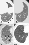
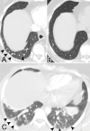
Results: Of the 48 patients, 45 patients required admission, with 18 admitted to the intensive care unit, and 13 requiring intubation. Seven patients (15%) were found to have pulmonary emboli. Dilated vessels were seen in 41 cases (85%), with 38 (78%) and 27 (55%) cases demonstrating vessel enlargement within and outside of lung opacities, respectively. Dilated distal vessels extending to the pleura and fissures were seen in 40 cases (82%) and 30 cases (61%), respectively. At DECT, mosaic perfusion pattern was observed in 24 cases (96%), regional hyperemia overlapping with areas of pulmonary opacities or immediately surrounding the opacities were seen in 13 cases (52%), opacities associated with corresponding oligemia were seen in 24 cases (96%), and hyperemic halo was seen in 9 cases (36%).
Conclusion: Pulmonary vascular abnormalities such as vessel enlargement and regional mosaic perfusion patterns are common in COVID-19 pneumonia. Perfusion abnormalities are also frequently observed at DECT in COVID-19 pneumonia and may suggest an underlying vascular process.Supplemental material is available for this article.© RSNA, 2020.
2020 by the Radiological Society of North America, Inc.
Conflict of interest statement
Disclosures of Conflicts of Interest: M.L. disclosed no relevant relationships. A.S. disclosed no relevant relationships. D.C. disclosed no relevant relationships. N.R. disclosed no relevant relationships. D.P.M. disclosed no relevant relationships. E.J.F. Activities related to the present article: disclosed no relevant relationships. Activities not related to the present article: disclosed grant funding paid to author from the American College of Radiology Innovation Fund and the National Cancer Institute Research Diversity Supplement for work not related to this manuscript. Other relationships: disclosed no relevant relationships. M.D.L. disclosed no relevant relationships. J.O.S. disclosed no relevant relationships. B.P.L. Activities related to the present article: disclosed no relevant relationships. Activities not related to the present article: disclosed money paid to author from Elsevier for textbook author and editor royalties. Other relationships: disclosed no relevant relationships.
Figures


References
-
- Li Y, Xia L. Coronavirus Disease 2019 (COVID-19): Role of Chest CT in Diagnosis and Management. AJR Am J Roentgenol 2020;214(6):1280–1286. - PubMed
-
- Zhao W, Zhong Z, Xie X, Yu Q, Liu J. Relation Between Chest CT Findings and Clinical Conditions of Coronavirus Disease (COVID-19) Pneumonia: A Multicenter Study. AJR Am J Roentgenol 2020;214(5):1072–1077. - PubMed
LinkOut - more resources
Full Text Sources
Medical
Miscellaneous

