. 2021 Jul;24(1):524.
doi: 10.3892/mmr.2021.12163. Epub 2021 May 26.

Effects of Txk‑mediated activation of NF‑κB signaling pathway on neurological deficit and oxidative stress after ischemia‑reperfusion in rats

Affiliations

Effects of Txk‑mediated activation of NF‑κB signaling pathway on neurological deficit and oxidative stress after ischemia‑reperfusion in rats

Qian-Lan Xu et al. Mol Med Rep. 2021 Jul.

Abstract

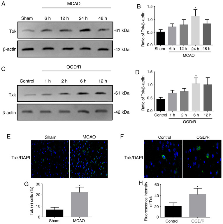
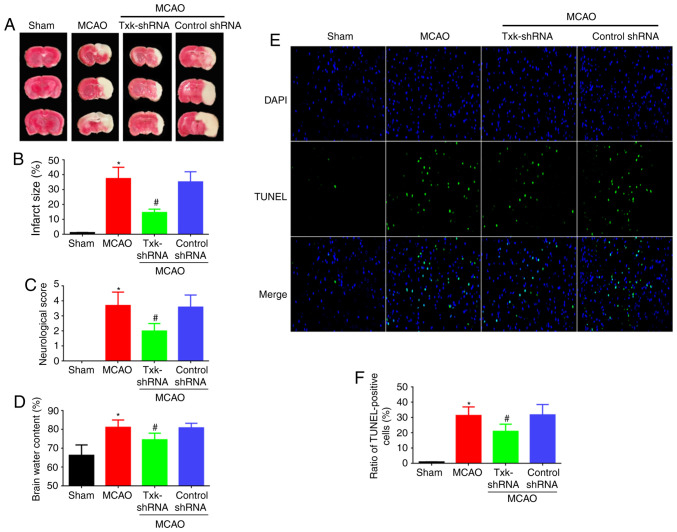
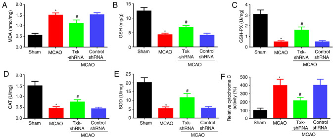
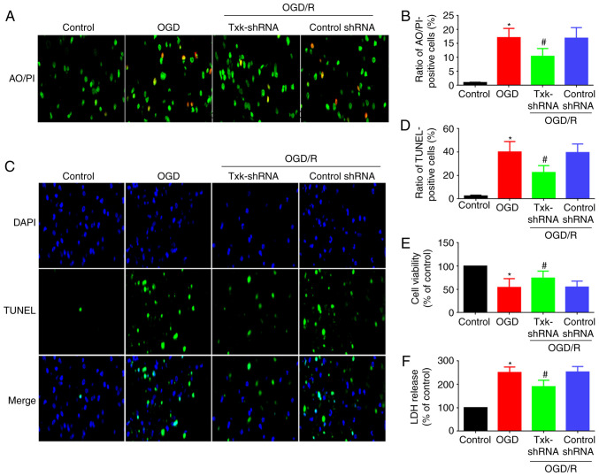
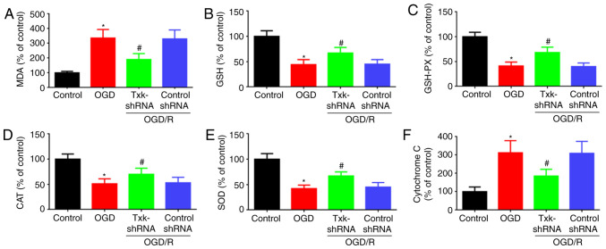
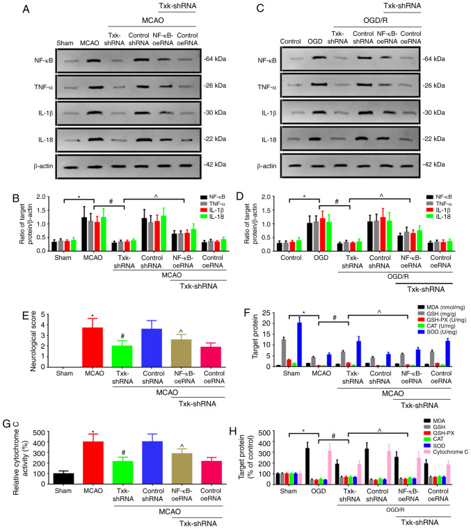
Ischemic stroke is an extremely mortal cerebrovascular disease, and neuroinflammation and oxidative stress emerge as important traits of ischemic stroke. However, as an inflammation‑associated factor, Txk tyrosine kinases (Txk) has been poorly studied in neuroscience research. The aim of the present study was to investigate the role of Txk after ischemia‑reperfusion (I/R) in vivo and in vitro, observe the association between Txk knockdown and neurological deficit and oxidative stress, and to explore whether the process was mediated by the activation of nuclear factor (NF)‑κB signaling pathway. Middle cerebral artery occlusion (MCAO), oxygen and glucose deprivation/reperfusion (OGD/R) model and western blotting have been used to simulate the I/R injury to analyze the expression, and to approximate the localization of Txk, respectively. Brain infarct volume, neurological score, brain water content, apoptosis and oxidative stress assays in vivo and apoptosis, cellular viability, the LDH release and oxidative stress assays in vitro were observed using a Txk‑knockdown lentivirus. Finally, NF‑κB overexpression lentivirus was applied to discuss whether the role of Txk following I/R was regulated by the NF‑κB signaling pathway. The results show that the Txk expression peaked at 24 h after MCAO and 6 h after OGD/R, respectively. Txk molecules gradually entered the nucleus after MCAO and OGD/R. The Txk‑knockdown lentivirus resulted in decreased brain infarct volume, neurological score, brain water content, apoptosis and oxidative stress after MCAO in vivo. Besides, Txk knockdown decreased apoptosis, LDH release, oxidative stress, and increased cellular viability, after ODG in vitro. Finally, NF‑κB overexpression reversed the process of neurological deficit and oxidative stress after Txk regulation in vivo and vitro. Overall, the present study suggests that Txk potentially regulates apoptosis, neurological deficit, and oxidative stress after I/R, by entering the nucleus. NF‑κB maybe the downstream target factor of Txk.

Keywords: Txk; ischemia‑reperfusion; neurological deficit; nuclear factor‑κB; oxidative stress.


Conflict of interest statement

The authors declare that they have no competing interests.

Figures

Figure 1.
Figure 1.
Western blotting and immunofluorescence results. (A) Western blot analysis of Txk expression in sham and MCAO 6-, 12-, 24- and 48-h groups. (B) Quantification of Txk in different groups. (C) Western blot analysis of Txk expression in control and OGD/R 1-, 2-, 6- and 12-h neurons. (D) Quantification of Txk in different neurons. (E) Immunofluorescence assay of Txk in sham and MCAO rats. Magnification, ×400) (F) Immunofluorescence assay of Txk in control and OGD/R neurons. Magnification, ×1000 (G) Txk-positive cells in sham and MCAO rats and (H) fluorescence intensity in control and OGD/R neurons. Protein level was normalized to β-actin. *P<0.05, MCAO 24 h vs. sham, MCAO 6, 12 and 48 h groups; OGD/R 6 h vs. control, OGD/R 1 and 2 h groups; MCAO vs. sham; OGD/R vs. control; n=6 per group. All data are represented as mean ± standard error. Txk, Txk tyrosine kinase; MCAO, middle cerebral artery occlusion; OGD/R, oxygen and glucose deprivation/reperfusion.
Figure 2.
Figure 2.
(A) 2,3,5-Triphenyltetrazolium chloride staining, (B) brain infarct volume assay, (C) neurological score assay, (D) brain water content, (E) immunofluorescence assay of TUNEL (magnification, ×400) and (F) TUNEL assay of sham, MCAO, Txk-shRNA and control-shRNA groups. *P<0.05, MCAO vs. sham group; #P<0.05, Txk-shRNA vs. MCAO group; n=6 per group. Txk, Txk tyrosine kinase; MCAO, middle cerebral artery occlusion; shRNA, short hairpin RNA.
Figure 3.
Figure 3.
(A) MDA, (B) GSH, (C) GSH-PX, (D) CAT, (E) SOD and (F) cytochrome C activity assays of sham, MCAO, Txk-shRNA and control-shRNA groups. *P<0.05, MCAO vs. sham group; #P<0.05, Txk-shRNA vs. MCAO group; n=6 per group. MCAO, middle cerebral artery occlusion; MDA, malondialdehyde; GSH, glutathione; GSH-PX, GSH peroxidase; CAT, catalase; SOD, superoxide dismutase; shRNA, short hairpin RNA.
Figure 4.
Figure 4.
(A) Immunofluorescence assay of AO/PI (magnification, ×400), (B) AO/PI-positive cells, (C) immunofluorescence assay of TUNEL (magnification, ×400), (D) TUNEL-positive cells (E) cellular viability and (F) LDH release assays of control, OGD/R, Txk-shRNA and control-shRNA groups. *P<0.05, OGD/R vs. control group; #P<0.05, Txk-shRNA vs. OGD/R group; n=6 per group. AO/PI, acridine orange/propidium iodide; Txk, Txk tyrosine kinase; MCAO, middle cerebral artery occlusion; OGD/R, oxygen and glucose deprivation/reperfusion; shRNA, short hairpin RNA.
Figure 5.
Figure 5.
(A) MDA, (B) GSH, (C) GSH-PX, (D) CAT, (E) SOD and (F) cytochrome C activity assays of control, OGD/R, Txk-shRNA and control-shRNA groups. *P<0.05, OGD/R vs. control group; #P<0.05, Txk-shRNA vs. OGD/R group; n=6 per group. MDA, malondialdehyde; GSH, glutathione; GSH-PX, GSH peroxidase; CAT, catalase; SOD, superoxide dismutase; Txk, Txk tyrosine kinase; shRNA, short hairpin RNA; OGD/R, oxygen and glucose deprivation/reperfusion.
Figure 6.
Figure 6.
(A) Western blot analysis and (B) quantification of NF-κB, TNF-α, IL-1β and IL-18 expression in sham, MCAO, Txk-shRNA, control shRNA, Txk-shRNA+NF-κB-oeRNA and Txk-shRNA+control oeRNA groups in vivo. (C) Western blot analysis and (D) quantification of NF-κB, TNF-α, IL-1β and IL-18 expression in control, OGD/R, Txk-shRNA, control shRNA, Txk-shRNA+NF-κB-oeRNA and Txk-shRNA+control oeRNA groups in vitro. (E) Neurological score assay, (F) MDA, GSH, GSH-PX, CAT and SOD assays and (G) cytochrome C activity assay in vivo. (H) MDA, GSH, GSH-PX, CAT, SOD and cytochrome C activity assays in vitro. Protein level was normalized to β-actin. *P<0.05, MCAO or OGD/R vs. sham or control group; #P<0.05, Txk-shRNA vs. MCAO or OGD/R group; ^P<0.05, Txk-shRNA+NF-κB-oeRNA vs. Txk-shRNA group; n=6 per group. NF-κB, nuclear factor-κB; TNF-α, tumor necrosis factor-α; IL, interleukin; MCAO, middle cerebral artery occlusion; OGD/R, oxygen and glucose deprivation/reperfusion; MDA, malondialdehyde; GSH, glutathione; GSH-PX, GSH peroxidase; CAT, catalase; SOD, superoxide dismutase; shRNA, short hairpin RNA; oe, overexpression.

References

    1. Kim JH, Nagy Á, Putzu A, Belletti A, Biondi-Zoccai G, Likhvantsev VV, Yavorovskiy AG, Landoni G. Therapeutic hypothermia in Critically Ill patients: A systematic review and meta-analysis of high quality randomized trials. Crit Care Med. 2020;48:1047–1054. - PubMed
    1. Wang J, Mao J, Wang R, Li S, Wu B, Yuan Y. Kaempferol protects against cerebral ischemia reperfusion injury through intervening oxidative and inflammatory stress induced apoptosis. Front Pharmacol. 2020;11:424. doi: 10.3389/fphar.2020.00424. - DOI - PMC - PubMed
    1. Boese AC, Lee JP, Hamblin MH. Neurovascular protection by peroxisome proliferator-activated receptor alpha in ischemic stroke. Exp Neurol. 2020;331:113323. doi: 10.1016/j.expneurol.2020.113323. - DOI - PubMed
    1. Ren Z, Zhang R, Li Y, Li Y, Yang Z, Yang H. Ferulic acid exerts neuroprotective effects against cerebral ischemia/reperfusion-induced injury via antioxidant and anti-apoptotic mechanisms in vitro and in vivo. Int J Mol Med. 2017;40:1444–1456. doi: 10.3892/ijmm.2017.3127. - DOI - PMC - PubMed
    1. Granger DN, Kvietys PR. Reperfusion injury and reactive oxygen species: The evolution of a concept. Redox Biol. 2015;6:524–551. doi: 10.1016/j.redox.2015.08.020. - DOI - PMC - PubMed

MeSH terms