Review
. 2022 Mar;36(7-9):367-388.
doi: 10.1089/ars.2021.0068. Epub 2021 Jul 7.

Redox Control of Integrin-Mediated Hepatic Inflammation in Systemic Autoimmunity

Affiliations
Review

Redox Control of Integrin-Mediated Hepatic Inflammation in Systemic Autoimmunity

Akshay Patel et al. Antioxid Redox Signal. 2022 Mar.

Abstract

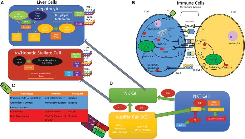
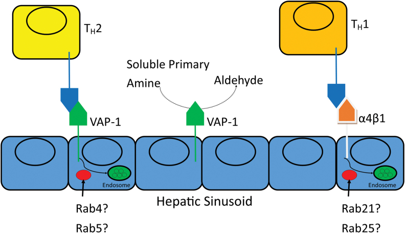
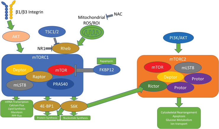
Significance: Systemic autoimmunity affects 3%-5% of the population worldwide. Systemic lupus erythematosus (SLE) is a prototypical form of such condition, which affects 20-150 of 100,000 people globally. Liver dysfunction, defined by increased immune cell infiltration into the hepatic parenchyma, is an understudied manifestation that affects up to 20% of SLE patients. Autoimmunity in SLE involves proinflammatory lineage specification in the immune system that occurs with oxidative stress and profound changes in cellular metabolism. As the primary metabolic organ of the body, the liver is uniquely capable to encounter oxidative stress through first-pass derivatization and filtering of waste products. Recent Advances: The traffic of immune cells from their development through recirculation in the liver is guided by cell adhesion molecules (CAMs) and integrins, cell surface proteins that tightly anchor cells together. The surface expression of CAMs and integrins is regulated via endocytic traffic that is sensitive to oxidative stress. Reactive oxygen species (ROS) that elicit oxidative stress in the liver may originate from the mitochondria, the cytosol, or the cell membrane. Critical Issues: While hepatic ROS production is a source of vulnerability, it also modulates the development and function of the immune system. In turn, the liver employs antioxidant defense mechanisms to protect itself from damage that can be harnessed to serve as therapeutic mechanisms against autoimmunity, inflammation, and development of hepatocellular carcinoma. Future Directions: This review is aimed at delineating redox control of integrin signaling in the liver and checkpoints of regulatory impact that can be targeted for treatment of inflammation in systemic autoimmunity. Antioxid. Redox Signal. 36, 367-388.

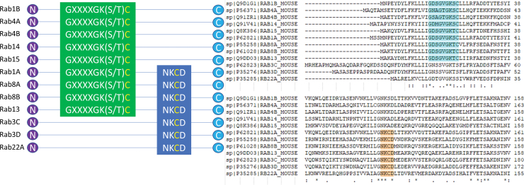
Keywords: Rab GTPases; T cell signaling; autoimmunity; cell adhesion molecules; endosomes; hepatic injury; immune synapse; immunity; integrins; liver; oxidative stress; reactive oxygen species; systemic lupus erythematosus.


Conflict of interest statement

The authors do not have any competing financial interests to disclose.

Figures

FIG. 1.
FIG. 1.
Effectors and targets of autoimmunity in the liver. (A) Rab GTPases regulate lipid droplet maturation, MPR trafficking to the trans Golgi network, and activate mTOR in hepatocytes. Ito/HSCs are responsible for vitamin A metabolism and fibrosis, which is regulated by Rab18. Rab18 promotes fibrosis/autophagy/lipophagy in response to liver injury. α1β1 (VLA-1) expression on hepatocyte and HSC surface is regulated by clathrin-dependent endocytosis and Dab2, but the involvement of Rab GTPases is unknown. α2β1 (VLA-2) expression on hepatocyte and HSC surface is regulated by Rab11. α5β1 (VLA-5) expression on HSC surface is regulated by Rab11 and the ROS-sensitive Rab25. (B) The interaction between a T cell and its cognate APC is tightly regulated. The MHC II/TCR interaction, CD2/CD58, and CD28/CD80 is the c-SMAC, while the interactions flanking this is the p-SMAC. Rab GTPases recycle the transmembrane proteins involved in the immune synapse. (C) A simple diagram of liver function within its context as a “filter” of sorts. The liver acts as a filter, detoxifier, and reducer of oxidative stress. Nutrients, processed foodstuffs, and bile pass through the portal triad, composed of the hepatic artery, the bile duct, and the portal vein. The major cells of the liver—hepatocyte, immune, and structural—are presented. Physiological functions are represented in blue, while disease pathologies are represented in red. After modification by biochemical mechanisms in the liver, processed materials are sent out to the rest of the body into the inferior vena cava, which drains into the right atrium of the heart. (D) NK cells, NKT cells, and Kupffer cells all act on each other and with other cells of the liver. NKT cells can promote hepatic inflammation through IFN-γ signaling when they are NKT I. NKT II reduces inflammation by activating cDCs, which suppress NKT I activity, thus reducing inflammatory liver dysfunction. NKT cells also promote NK cell signaling through the release of IFN-γ. NK cells can inhibit Ito/HSC signaling and hepatocyte signaling. APC, antigen presenting cell; CD, cluster of differentiation; cDC, conventional dendritic cell; c-SMAC, central supramolecular activation cluster; Dab2, disabled homolog 2; ER, endoplasmic reticulum; HSC, hepatic stellate cell; ICAM-1, intracellular adhesion molecule-1; IFN-γ, interferon gamma; IL-12/IL-18, interleukin 12/18; LFA-1, lymphocyte function-associated antigen 1; MHC II, major histocompatibility complex II; MPR, mannose-6-phosphate receptor; mTOR, mechanistic target of rapamycin; NK, natural killer; NKT, natural killer T; NKT I, type I NKT; NKT II, type II NKT; p-SMAC, peripheral supramolecular activation cluster; Rab, Ras-like protein from rat brain; ROS, reactive oxygen species; TCR, T cell receptor; VLA-1, very late antigen-1; integrin α1β1; VLA-2, very late antigen-2; integrin α2β1; VLA-5, very late antigen-5; integrin α5β1; fibronectin receptor. Color images are available online.
FIG. 3.
FIG. 3.
VAP-1 recruits leukocytes and oxidizes primary amines in the liver during inflammatory states. VAP-1 is a cell surface protein with enzymatic activity, which binds to CD11b to recruit macrophages that may mediate liver damage in autoimmunity. Studies using VAP-1-GFP fusion proteins localized VAP-1 to early endosomes, suggesting that Rab GTPases such as Rab4 and Rab5 may be involved in the cell surface expression of VAP-1. VAP-1, vascular adhesion protein. Color images are available online.
FIG. 2.
FIG. 2.
Redox-sensitive motifs in Rab GTPases can control cell surface protein expression. GTP, redox agents, or both can allosterically activate GTPases depending on binding motifs. Representative schematics of 12 mouse Rab GTPases, which contain either the GXXXGK(S/T)C motif, the NKCD motif, or both. A Clustal Omega alignment is provided for reference, with the GXXXGK(S/T)C motif in blue and the NKCD motif in orange. Aligned Rab GTPases, sensitive to ROS, can mediate integrin and cell surface protein expression relevant to SLE and autoimmunity. GTP, guanosine triphosphate; SLE, systemic lupus erythematosus. Color images are available online.
FIG. 4.
FIG. 4.
The mTOR pathway responds to oxidative stress and integrin signals. While both mTORC1 and mTORC2 respond to rapamycin in an inhibitory manner, mTORC2 promotes downstream cellular growth effects through upstream PI3K/AKT signaling. mTORC1 responds to Rheb through ROS, TSC1/2, and the newly discovered drug NR1 to mediate downstream effects. AKT also promotes mTORC1 activity through β1 and β3 signaling. mTORC1, complex 1 of the mechanistic target of rapamycin; PI3K, phosphatidylinositol 3-kinase; Rheb, Ras homolog enriched in brain; ROI, reactive oxygen intermediate; TSC, tuberous sclerosis complex. Color images are available online.
FIG. 5.
FIG. 5.
Current antioxidant therapies and their potential in reversing liver dysfunction. Sorbinil inhibits aldose reductase to prevent the catalysis of NADPH, resulting in increased NADPH concentrations. Vitamins C and E act through ascorbate reductase and Vit E radical reductase to transfer electrons from ROS-damaged proteins to glutathione. NAC, a potent antioxidant, is a precursor to GSH through a cysteine intermediate. NAC reduces glutathione depletion, ROS generation, and potentially VAP-1 mediated T cell recruitment as a result of liver damage that can occur from oxidative stress, ethanol intoxication, HBV or HCV infection. Metadoxine and Silymarin/Silybin regenerate glutathione peroxidase to facilitate ROS clearance through a glutathione-dependent mechanism. GSH, reduced glutathione; HBV, hepatitis B virus; HCV, hepatitis C virus; IV, intravenous; NAC, N-acetylcysteine; NADPH, nicotinamide adenine dinucleotide phosphate; NAI-ALF, nonacetaminophen-induced acute liver failure; Vit C, vitamin C; Vit E, vitamin E. Color images are available online.
FIG. 6.
FIG. 6.
Antioxidant pathways in the liver. The redox pathways present in the cytosol and the mitochondria act to reduce oxidative stress by generating NADPH, glutathione, or directly reducing harmful ROS into their neutral counterparts. The box around the transaldolase and G6PD reactions represents the interaction between the oxidative and nonoxidative branches of the pentose phosphate pathway linked by PGI. The thioredoxin proteins converge to facilitate Prx activity; this is detailed in Figure 7. 6PGL, 6-phosphoglucono-δ-lactone; E4P, erythrose 4-phosphate; F6P, fructose 6-phosphate; G6P, glucose 6-phosphate; G6PD, glucose-6-phosphate dehydrogenase; GA3P, glyceraldehyde 3-phosphate; GSSG, oxidized glutathione; IDH2, isocitrate dehydrogenase 2; NADH, nicotinamide adenine dinucleotide; NNT, nicotinamide nucleotide transhydrogenase; PGI, phosphoglucoisomerase; Prx, peroxiredoxin; S7P, sedoheptulose 7-phosphate. Color images are available online.
FIG. 7.
FIG. 7.
The Prxs, which reduce oxidative stress, operated via three variant mechanisms based on the number of cysteines involved and their localization within two (typical 2-Cys formation) or a single subunit molecules (atypical 2-Cys formation) or reliance of a single cysteine (1-Cys formation). Cys, cysteine; Grx, glutaredoxin. Color images are available online.

Similar articles

Cited by

References

    1. Andres PG, Howland KC, Dresnek D, Edmondson S, Abbas AK, and Krummel MF. CD28 signals in the immature immunological synapse. J Immunol 172: 5880–5886, 2004. - PubMed
    1. Bai X, Wang J, Guo Y, Pan J, Yang Q, Zhang M, Li H, Zhang L, Ma J, Shi F, Shu W, Wang Y, and Leng J. Prostaglandin E2 stimulates β1-integrin expression in hepatocellular carcinoma through the EP1 receptor/PKC/NF-κB pathway. Sci Rep 4: 6538, 2014. - PMC - PubMed
    1. Ballatori N, Krance SM, Notenboom S, Shi S, Tieu K, and Hammond CL. Glutathione dysregulation and the etiology and progression of human diseases. Biol Chem 390: 191–214, 2009. - PMC - PubMed
    1. Banales JM, Huebert RC, Karlsen T, Strazzabosco M, LaRusso NF, and Gores GJ. Cholangiocyte pathobiology. Nat Rev Gastroenterol Hepatol 16: 269–281, 2019. - PMC - PubMed
    1. Bandyopadhyay K, Marrero I, and Kumar V. NKT cell subsets as key participants in liver physiology and pathology. Cell Mol Immunol 13: 337–346, 2016. - PMC - PubMed

Publication types

LinkOut - more resources