

Contrast-Enhanced Ultrasound Imaging of Uterine Disorders: A Systematic Review
- PMID: 34036872
- PMCID: PMC8299780
- DOI: 10.1177/01617346211017462
Contrast-Enhanced Ultrasound Imaging of Uterine Disorders: A Systematic Review
Abstract
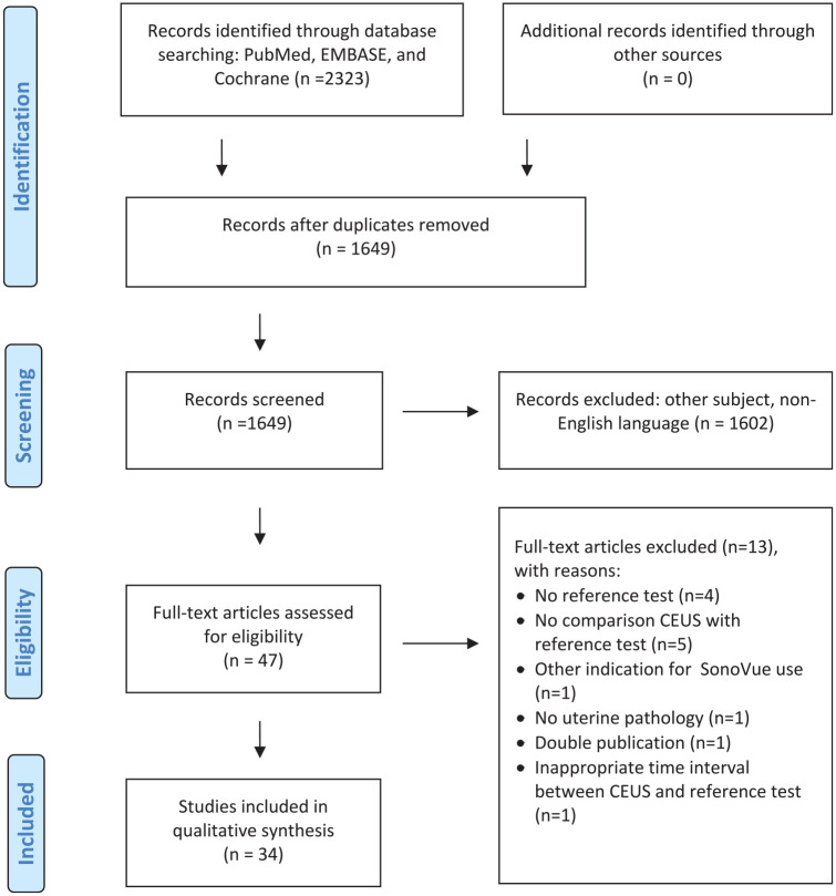
Uterine disorders are often presented with overlapping symptoms. The microvasculature holds specific information important for diagnosing uterine disorders. Conventional sonography is an established diagnostic technique in gynecology, but is limited by its inability to image the microvasculature. Contrast-enhanced ultrasound (CEUS), is capable of imaging the microvasculature by means of intravascular contrast agents; that is, gas-filled microbubbles. We provide a literature overview on the use of CEUS in diagnosing myometrial and endometrial disorders, that is, fibroids, adenomyosis, leiomyosarcomas and endometrial carcinomas, as well as for monitoring and enhancing the effectiveness of minimally invasive therapies. A systematic literature search with quality assessment was performed until December 2020. In total 34 studies were included, published between 2007 and 2020.The results entail a description of contrast-enhancement patterns obtained from healthy tissue and from malignant and benign tissue; providing a first base for potential diagnostic differentiation in gynecology. In addition it is also possible to determine the degree of myometrial invasion in case of endometrial carcinoma using CEUS. The effectiveness of minimally invasive therapies for uterine disorders can safely and accurately be assessed with CEUS. In conclusion, the abovementioned applications of CEUS are promising and it is worth further exploring its full potential for gynecology by designing innovative and methodologically high-quality clinical studies.
Keywords: contrast-enhanced ultrasound; imaging; microvasculature; systematic review; uterine disorders.
Conflict of interest statement
Figures
Similar articles
-
Use of Contrast-Enhanced Ultrasound in the Assessment of Uterine Fibroids: A Feasibility Study.Ultrasound Med Biol. 2018 Aug;44(8):1901-1909. doi: 10.1016/j.ultrasmedbio.2018.03.030. Epub 2018 May 4. Ultrasound Med Biol. 2018. PMID: 29735316
-
Intraprocedure contrast enhanced ultrasound: the value in assessing the effect of ultrasound-guided high intensity focused ultrasound ablation for uterine fibroids.Ultrasonics. 2015 Apr;58:123-8. doi: 10.1016/j.ultras.2015.01.005. Epub 2015 Jan 17. Ultrasonics. 2015. PMID: 25627929
-
Therapeutic response assessment of high intensity focused ultrasound therapy for uterine fibroid: utility of contrast-enhanced ultrasonography.Eur J Radiol. 2007 May;62(2):289-94. doi: 10.1016/j.ejrad.2006.11.039. Epub 2007 Jan 26. Eur J Radiol. 2007. PMID: 17258417
-
Contrast-Enhanced Ultrasound for Monitoring Non-surgical Treatments of Uterine Fibroids: A Systematic Review.Ultrasound Med Biol. 2021 Jan;47(1):3-18. doi: 10.1016/j.ultrasmedbio.2020.09.016. Epub 2020 Oct 22. Ultrasound Med Biol. 2021. PMID: 33239156 Free PMC article.
-
A Systematic Review on the Use of Qualitative and Quantitative Contrast-enhanced Ultrasound in Diagnosing Testicular Abnormalities.Urology. 2021 Aug;154:16-23. doi: 10.1016/j.urology.2021.02.012. Epub 2021 Feb 20. Urology. 2021. PMID: 33621585
Cited by
-
Comparison of the diagnostic accuracy of contrast-enhanced/DWI MRI and ultrasonography in the differentiation between benign and malignant myometrial tumors.Ann Med Surg (Lond). 2021 Sep 21;70:102813. doi: 10.1016/j.amsu.2021.102813. eCollection 2021 Oct. Ann Med Surg (Lond). 2021. PMID: 34691413 Free PMC article.
-
Applying contrast-enhanced ultrasound model to distinguish atypical focal adenomyosis from uterine leiomyomas.Ann Transl Med. 2022 Oct;10(20):1108. doi: 10.21037/atm-22-4460. Ann Transl Med. 2022. PMID: 36388773 Free PMC article.
-
Diagnostic value of transvaginal contrast-enhanced ultrasound in identifying benign and malignant endometrial lesions and assessing myometrial invasion.Ultrasonography. 2024 Nov;43(6):448-456. doi: 10.14366/usg.24097. Epub 2024 Jul 20. Ultrasonography. 2024. PMID: 39327718 Free PMC article.
-
Clinical value of quantitative analysis of contrast-enhanced ultrasonography in the differential diagnosis of benign and malignant pelvic tumors.Quant Imaging Med Surg. 2023 Oct 1;13(10):6636-6645. doi: 10.21037/qims-23-582. Epub 2023 Aug 24. Quant Imaging Med Surg. 2023. PMID: 37869279 Free PMC article.
-
Value of contrast-enhanced ultrasonography in microwave ablation treatment of symptomatic focal uterine adenomyosis.Ultrasonography. 2024 Jan;43(1):68-77. doi: 10.14366/usg.23145. Epub 2023 Nov 7. Ultrasonography. 2024. PMID: 38109892 Free PMC article.
References
-
- Testa AC, Ferrandina G, Fruscella E, Van Holsbeke C, Ferrazzi E, Leone FP, et al. The use of contrasted transvaginal sonography in the diagnosis of gynecologic diseases: a preliminary study. J Ultrasound Med. 2005;24(9):1267-78. - PubMed
-
- Dijkmans PA, Juffermans LJ, Musters RJ, van Wamel A, ten Cate FJ, van Gilst W, et al. Microbubbles and ultrasound: from diagnosis to therapy. Eur J Echocardiogr. 2004;5(4):245-56. - PubMed
-
- Brannigan M, Burns PN, Wilson SR. Blood flow patterns in focal liver lesions at microbubble-enhanced US. Radiographics. 2004;24(4):921-35. - PubMed
-
- Exacoustos C, Romanini ME, Amadio A, Amoroso C, Szabolcs B, Zupi E, et al. Can gray-scale and color Doppler sonography differentiate between uterine leiomyosarcoma and leiomyoma? J Clin Ultrasound. 2007;35(8):449-57. - PubMed
-
- Zhang XL, Zheng RQ, Yang YB, Huang DM, Song Q, Mao YJ, et al. The use of contrast-enhanced ultrasound in uterine leiomyomas. Chin Med J (Engl). 2010;123(21):3095-9. - PubMed
Publication types
MeSH terms
Substances
LinkOut - more resources
Full Text Sources
Other Literature Sources
