Review
. 2021 Jun 16;19(23):5048-5062.
doi: 10.1039/d1ob00608h.

Asparaginyl endopeptidases: enzymology, applications and limitations

Affiliations
Review

Asparaginyl endopeptidases: enzymology, applications and limitations

T M Simon Tang et al. Org Biomol Chem. .

Abstract

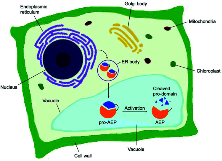
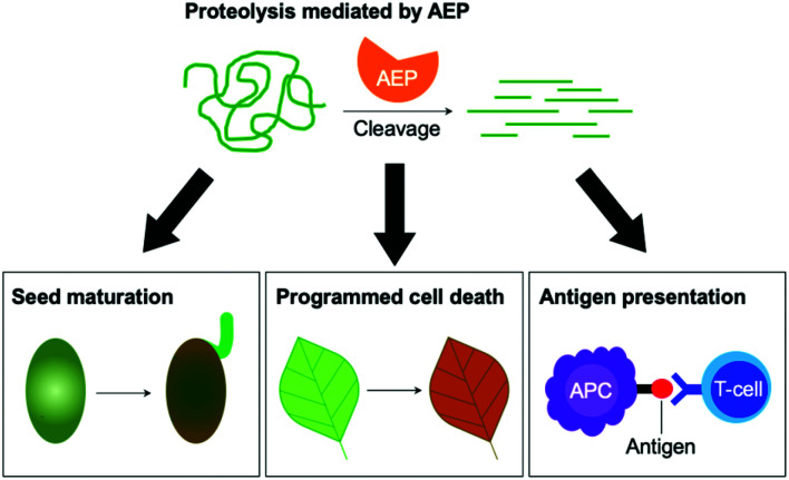
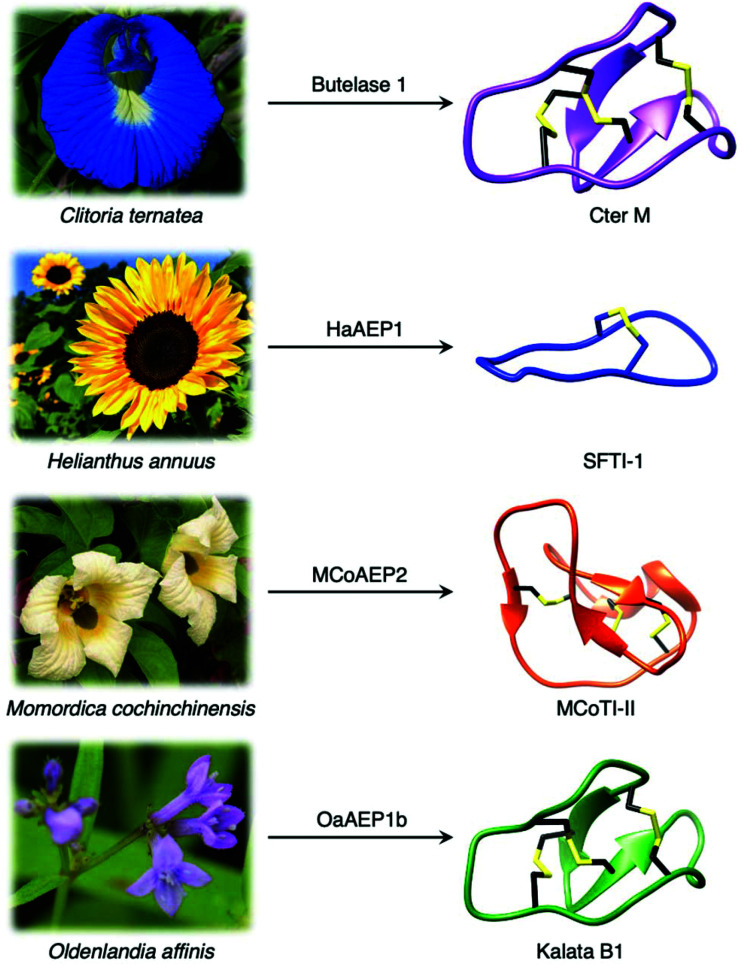
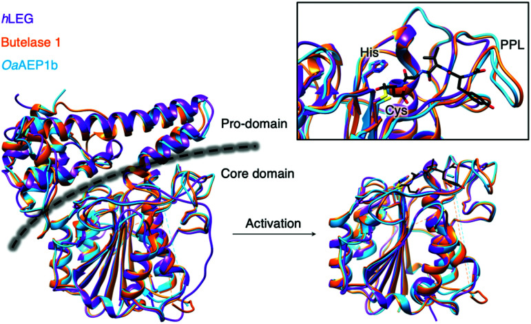
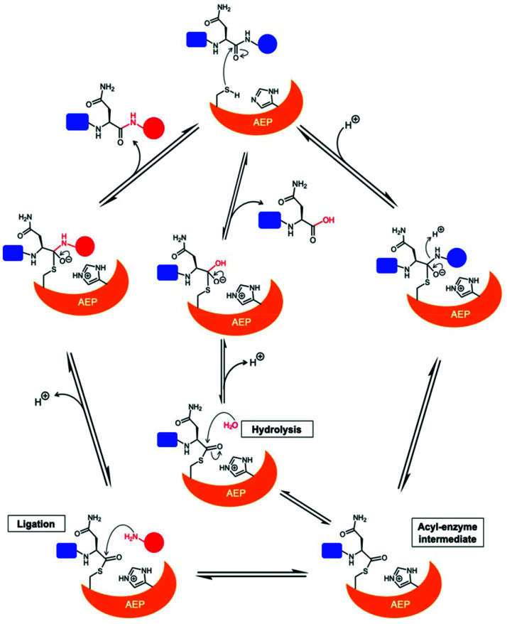
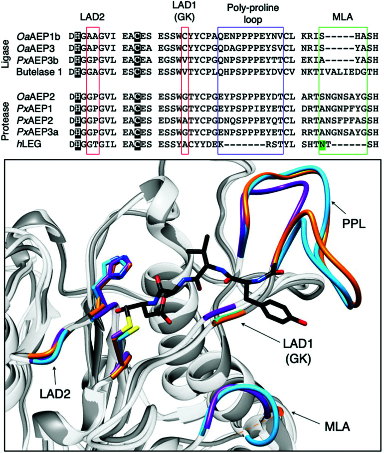
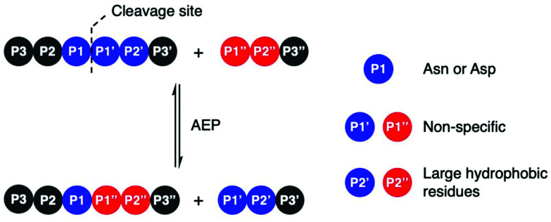
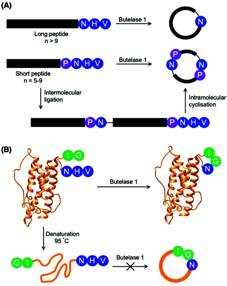
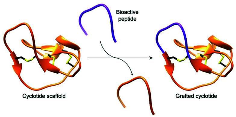
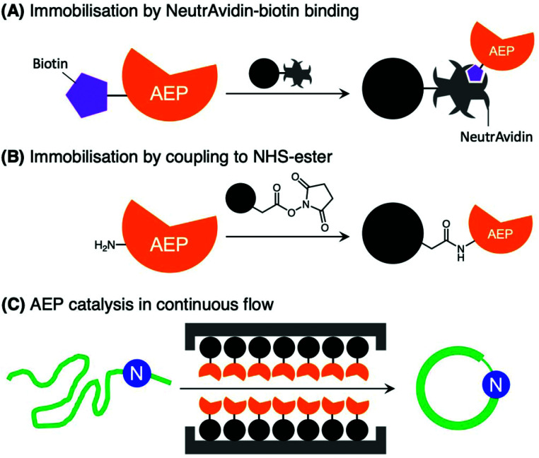
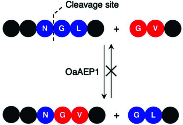
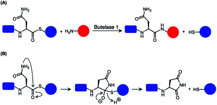
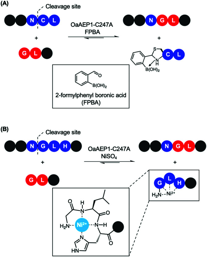
Asparaginyl endopeptidases (AEP) are cysteine proteases found in mammalian and plant cells. Several AEP isoforms from plant species were found to exhibit transpeptidase activity which is integral for the key head-to-tail cyclisation reaction during the biosynthesis of cyclotides. Since many plant AEPs exhibit excellent enzyme kinetics for peptide ligation via a relatively short substrate recognition sequence, they have become appealing tools for peptide and protein modification. In this review, research focused on the enzymology of AEPs and their applications in polypeptide cyclisation and labelling will be presented. Importantly, the limitations of using AEPs and opportunities for future research and innovation will also be discussed.


Conflict of interest statement

There are no conflicts to declare.

Figures

Fig. 1
Fig. 1. Biosynthesis of AEP in plant cells. Zymogenic AEPs are folded in the ER and stored in ER bodies. The pro-enzyme is trafficked to the vacuole, where the acidic environment enables cleavage of the pro-domain resulting in activation.
Fig. 2
Fig. 2. The peptidase activities of AEPs are involved in several critical cellular processes including seed maturation, programmed cell death and the regulation of antigen presentation.
Fig. 3
Fig. 3. Plant species utilise AEPs with ligase activities to generate backbone cyclised peptides with enhanced stability and bioactivity. Cter M (PDB: 2LAM) from C. ternatea, SFTI-1 (PDB: 1JBL) from H. annuus, MCoTI-II (PDB: 1IB9) from M. cochinchinensis and kalata B1 (PDB: 1NB1) from O. affinis.
Fig. 4
Fig. 4. Overlaid ribbon representations of human pro-legumain (hLEG, PDB: 4FGU, purple), butelase 1 (PDB: 6DHI, orange), and OaAEP1b (PDB: 5H0I, blue) structures deduced from X-ray crystallography. The black dotted line indicates the separation between the pro-domain and the core domain. After activation, modified butelase 1 (PDB: 6DHI) and OaAEP1b (PDB: 5H0I) structures showing only the catalytic core domains were overlaid with the crystal structure of activated human legumain (PDB: 4AWA, purple). In the box, an enlarged image of the active sites reveals the respective catalytic diads (Cys and His), the conformation adopted by the inhibitor (YVAD-CMK) in complex with activated human legumain, and a plant specific loop region referred to as the poly-proline loop (PPL).
Scheme 1
Scheme 1. Proposed mechanism of AEPs catalysis with a Cys/His catalytic diad. AEP recognises and cleaves a peptide substrate containing Asn to form an acyl-enzyme intermediate via a tetrahedral intermediate. A nucleophilic attack resolves the thioester intermediate to afford the product peptide and the AEP is regenerated. The nucleophile can be a water molecule or the N-terminus of a peptide, resulting in peptide hydrolysis or ligation, respectively.
Fig. 5
Fig. 5. Key features associated with the determination AEP ligase and protease activity. Multiple sequence alignment of AEPs, from O. affinis (OaAEP1b, 3), P. x hybrida (PxAEP1, 2, 3a, 3b), C. ternatea (butelase 1) and H. sapien (hLEG), centred around amino acid residues reported to influence activity. Catalytic diads (Cys/His) are shown in white letters with black highlight, N-glycosylation site is highlighted in green, the overlaid red boxes show LAD1 (gatekeeper/GK) and LAD2, blue box shows the plant-specific poly-proline loop and the green box highlights the MLA. In the box, an enlarged overlaid image of OaAEP1 (PDB: 5H0I), butelase 1 (PDB: 6DHI) and human legumain (PDB: 4AWA) active sites. Key features (catalytic diad, LAD1, LAD2, PPL and MLA) are rendered blue, orange and purple for OaAEP1, butelase 1, and human legumain respectively. In black, the conformation adopted by the inhibitor (YVAD-CMK) in complex with human legumain.
Scheme 2
Scheme 2. AEP-mediated transpeptidation, with amino acid residues numbered according to Schechter and Berger. The putative recognition sequences P1–P1′–P2′ and P1′′–P2′′ are coloured in blue and red, respectively.
Scheme 3
Scheme 3. Peptide cyclisation catalysed by butelase 1. (A) Peptides greater than 9 amino acid residues in length and bearing the butelase 1 substrate recognition sequence (NHV) are cyclised by the AEP. Whereas peptides of 5–9 amino acid residues in length, with Pro at P2 and bearing the butelase 1 substrate recognition sequence (NHV) are ligated, then cyclised by AEP to afford cyclic oligomers (only cyclo-dimer shown here). (B) Butelase 1 was reported to cyclise the folded somatropin (PDB: 1HGU) but not the denatured protein.
Fig. 6
Fig. 6. Cartoon representation of a grafted cyclotide. A bioactive peptide (purple) is inserted into a cyclotide backbone, shown here using the structure of MCoTI-II (PDB: 1IB9).
Fig. 7
Fig. 7. Immobilisation of AEPs on solid support. AEPs, butelase1 and VyPAL2, have been immobilised on agarose beads via (A) NeutrAvidin-biotin affinity binding and (B) direct coupling to N-hydroxysuccinimide (NHS) ester with primary amines presented by lysine residues. (C) The immobilised AEPs were reported to facilitate ligation of proteins and peptides in continuous flow.
Scheme 4
Scheme 4. Ligation of self-assembling protein domains by OaAEP1b. Pre-organisation of the ligating substrates bring the reactive sites into close proximity, which was shown to affect AEP catalysis.
Scheme 5
Scheme 5. OaAEP1 recognises Val at P2′′, but poorly at P2′. Consequently, this feature in the substrate recognition of OaAEP1 has been exploited to prevent the undesired reverse reaction.
Fig. 8
Fig. 8. Comparison of procedures for recombinant OaAEP1-C247A. (Red) Recombinant AEP preparation from the pro-enzyme involves multiple purification steps and an incubation period in acidic conditions (pH 4.0). (Green) Recombinant AEP preparation without the pro-domain enabled a streamlined method with less chromatographic steps and does not require activation. (IMAC) Immobilised metal affinity chromatography, (SAX) strong anion exchange and (SEC) size exclusion chromatography. X-ray crystal structure of OaAEP1b (PDB: 5H0I) used here as a representative AEP.
Scheme 6
Scheme 6. Use of thiodepsipeptide for butelase 1-mediated ligation. (A) Thioester linked Asn is accepted by butelase 1. Peptide ligation generates a peptide by-product with a α-thiol which is not recognised as a nucleophilic peptide substrate. As a result, the reaction was rendered irreversible. (B) Proposed mechanism for the hydrolysis of the unstable thioester.
Scheme 7
Scheme 7. Nucleophile quenching strategies to enable effective peptide ligation by OaAEP1-C247A. (A) FPBA reacts with N-terminal cysteine to thiazolidine derivative to prevent reverse reaction. (B) Divalent Ni2+ quenches the nucleophilic peptide by-product generated by OaAEP1-C247A, thus driving the reaction equilibrium towards product formation. The tripeptide motif Gly-Leu-His at the N-terminus coordinates to Ni2+.
None
T. M. Simon Tang (left) Louis Y. P. Luk (right)

Similar articles

Cited by

References

    1. Hara-Nishimura I. Takeuchi Y. Nishimura M. Molecular characterization of a vacuolar processing enzyme related to a putative cysteine proteinase of Schistosoma mansoni. Plant Cell. 1993;5:1651. - PMC - PubMed
    1. Chen J.-M. Dando P. M. Stevens R. A. E. Fortunato M. Barrett A. J. Cloning and expression of mouse legumain, a lysosomal endopeptidase. Biochem. J. 1998;335:111–117. - PMC - PubMed
    1. Dall E. Brandstetter H. Structure and function of legumain in health and disease. Biochimie. 2016;122:126–150. - PubMed
    1. Hemu X. El Sahili A. Hu S. Wong K. Chen Y. Wong Y. H. Zhang X. Serra A. Goh B. C. Darwis D. A. Chen M. W. Sze S. K. Liu C.-F. Lescar J. Tam J. P. Structural determinants for peptide-bond formation by asparaginyl ligases. Proc. Natl. Acad. Sci. U. S. A. 2019;116:11737–11746. - PMC - PubMed
    1. Hayashi Y. Yamada K. Shimada T. Matsushima R. Nishizawa N. K. Nishimura M. Hara-Nishimura I. A proteinase-storing body that prepares for cell death or stresses in the epidermal cells of Arabidopsis. Plant Cell Physiol. 2001;42:894–899. - PubMed

Publication types

MeSH terms

Substances

LinkOut - more resources