. 2021 May 24;54(8):e11073.
doi: 10.1590/1414-431X2020e11073. eCollection 2021.

Effects of vitamin D-induced supernatant of placental explants from preeclamptic women on oxidative stress and nitric oxide bioavailability in human umbilical vein endothelial cells

Affiliations

Effects of vitamin D-induced supernatant of placental explants from preeclamptic women on oxidative stress and nitric oxide bioavailability in human umbilical vein endothelial cells

P R Nunes et al. Braz J Med Biol Res. .

Abstract

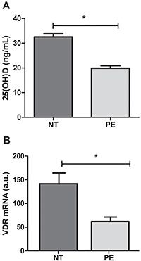
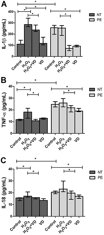
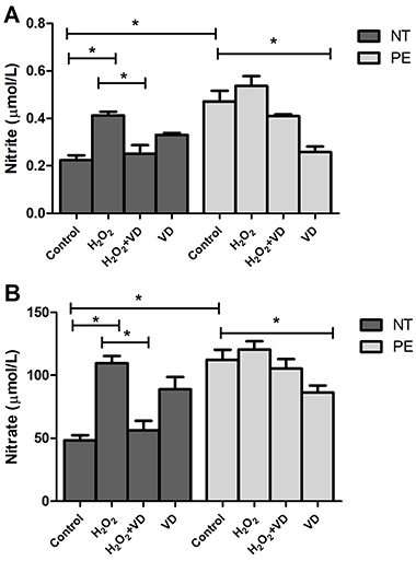
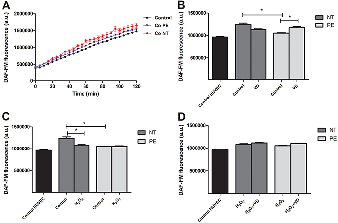
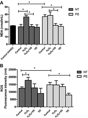
The study evaluated the effect of the supernatant of placental explants from preeclamptic (PE) and normotensive (NT) pregnant women after tissue treatment with or without vitamin D (VD) on oxidative stress and nitric oxide (NO) bioavailability in human umbilical vein endothelial cells (HUVEC). Placental explants were prepared from eight NT and eight PE women, and supernatants were obtained after incubation with or without hydrogen peroxide (H2O2) and/or VD. HUVEC were cultured for 24 h with supernatants, and the following parameters were analyzed in HUVEC cultures: NO, nitrate (NO3-), and nitrite (NO2-) levels, lipid peroxidation, and intracellular reactive oxygen species (ROS). Results showed that the production of NO3-, NO2-, malondialdehyde (MDA), and ROS were significantly higher in HUVEC treated with explant supernatant from PE compared to NT pregnant women, while the supernatant of PE explants treated with VD led to a decrease in these parameters. A significantly high production of NO was detected in HUVEC cultured with control supernatant of NT group, and in cultures treated with supernatant of PE explants treated with VD. Taken together, these results demonstrated that cultures of placental explants from PE women with VD treatment generated a supernatant that decreased oxidative stress and increased the bioavailability of NO in endothelial cells.


Figures

Figure 1
Figure 1. Levels of vitamin 25 (OH) D in plasma (A) and vitamin D receptor (VDR) gene expression (B) in the placental explants from preeclamptic (PE) (n=8) and normotensive (NT) pregnant women (n=8). Data are reported as means±SD. *P<0.05 (Mann-Whitney U test). a.u.: arbitrary units.
Figure 2
Figure 2. Concentration of cytokines in supernatant of placental explants from preeclamptic (PE) (n=8) and normotensive (NT) pregnant women (n=8) under different treatments. Data are reported as median with range. *P<0.05 (Mann-Whitney U test). IL-1β: interleukin-1 beta; TNF-α: tumor necrosis factor-alpha; IL-18: interleukin-18; VD: vitamin D.
Figure 3
Figure 3. Levels of nitrite (A) and nitrate (B) in the supernatant of human umbilical vein endothelial cells treated with supernatant of placental explants from preeclamptic (PE) (n=8) and normotensive (NT) pregnant women (n=8), cultured in the absence (Control) or presence of H2O2, H2O2+VD, and VD. Data are reported as means±SD for one independent experiment, performed in quintuplicates. *P<0.05 (ANOVA followed by Bonferroni's multiple comparison test). VD: vitamin D.
Figure 4
Figure 4. Nitric oxide fluorescence signal measured using DAF-FM™ after human umbilical vein endothelial cells (HUVEC) were exposed to placental explant supernatants from preeclamptic (PE) (n=8) and normotensive (NT) pregnant women (n=8). A, Control, Control NT, and Control PE; B, effect of treatment with vitamin D (VD) at 60 min; C, effect of treatment with H2O2 at 60 min; and D, effect of treatment with H2O2+VD at 60 min. Data are reported as means±SD for one independent experiment, performed in quintuplicates. *P<0.05 (ANOVA followed by Bonferroni's multiple comparison test). a.u.: arbitrary units.
Figure 5
Figure 5. Malondialdehyde (MDA) levels (A) and levels of intracellular reactive oxygen species (ROS) (B) evaluated by fluorescence of 2,7-dichlorodihydrofluorescein diacetate (DCFH-DA) in the supernatant of human umbilical vein endothelial cells (HUVEC) treated with supernatant of placental explants from preeclamptic (PE) (n=8) and normotensive (NT) pregnant women (n=8), cultured in the absence (Control) or presence of H2O2, H2O2+vitamin D (VD), and VD. Data are reported as means±SD. *P<0.05 (ANOVA followed by Bonferroni's multiple comparison test).

Similar articles

Cited by

References

    1. American College of Obstetricians and Gynecologists Clinical Management Guidelines for Obstetrician-Gynecologists. Acog practice bulletin No. 202: Gestational hypertension and preeclampsia. Obstet Gynecol. 2009;133:e1–e25.
    1. Folk DM. Hypertensive disorders of pregnancy: overview and current recommendations. J Midwifery Womens Health. 2018;63:289–300. doi: 10.1111/jmwh.12725. - DOI - PubMed
    1. Moncada S, Higgs A. The L-arginine nitric-oxide pathway. N Eng J Med. 1993;329:2002–2012. doi: 10.1056/NEJM199312303292706. - DOI - PubMed
    1. Krause BJ, Hanson MA, Casanello P. Role of nitric oxide in placental vascular development and function. Placenta. 2011;32:797–805. doi: 10.1016/j.placenta.2011.06.025. - DOI - PMC - PubMed
    1. Poniedzialek-Czajkowska E, Marciniak B, Kimber-Trojnar Z, Leszczyńska-Gorzelak B, Oleszczuket J. Nitric oxide in normal and preeclamptic pregnancy. Curr Pharm Biotechnol. 2011;12:743–749. doi: 10.2174/138920111795470921. - DOI - PubMed