. 2021 Jun 16;12(12):2167-2181.
doi: 10.1021/acschemneuro.1c00169. Epub 2021 May 26.

Specific Phospholipid Modulation by Muscarinic Signaling in a Rat Lesion Model of Alzheimer's Disease

Affiliations

Specific Phospholipid Modulation by Muscarinic Signaling in a Rat Lesion Model of Alzheimer's Disease

Alberto Llorente-Ovejero et al. ACS Chem Neurosci. .

Abstract

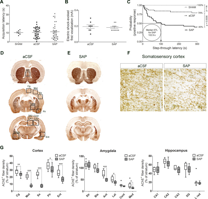
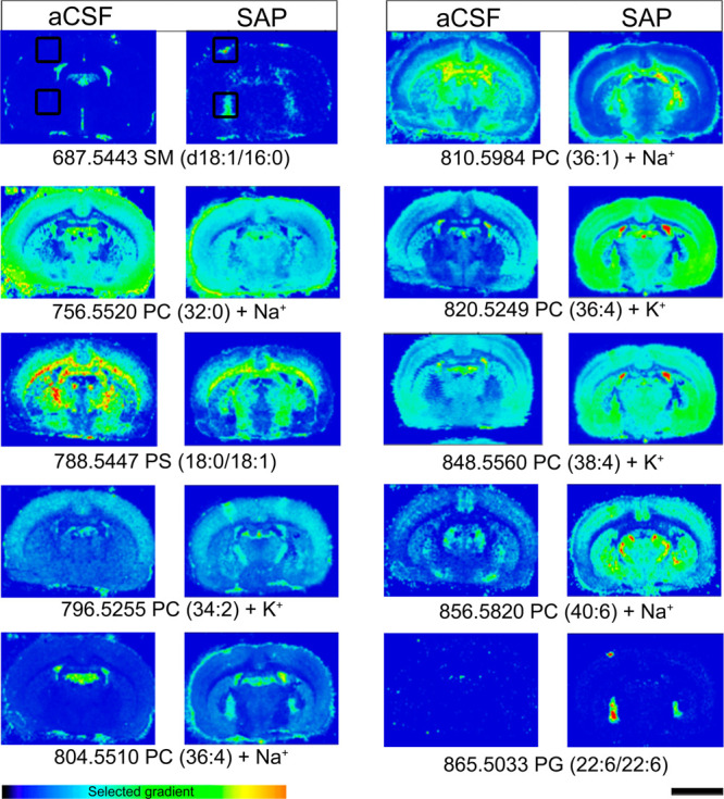
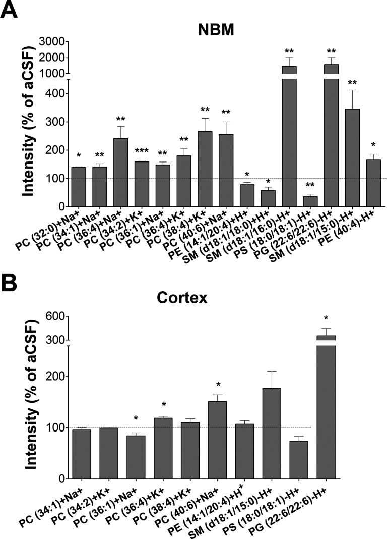
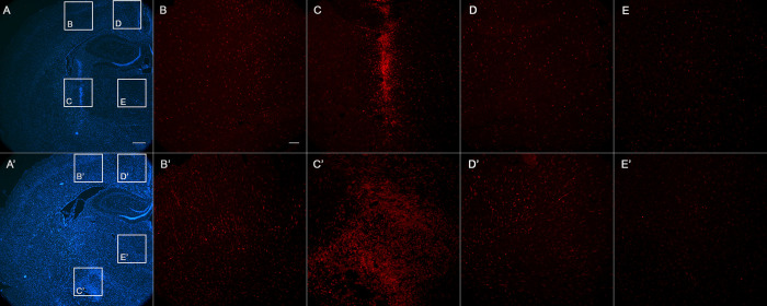
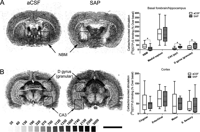
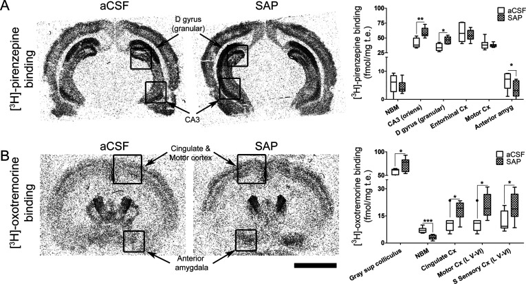
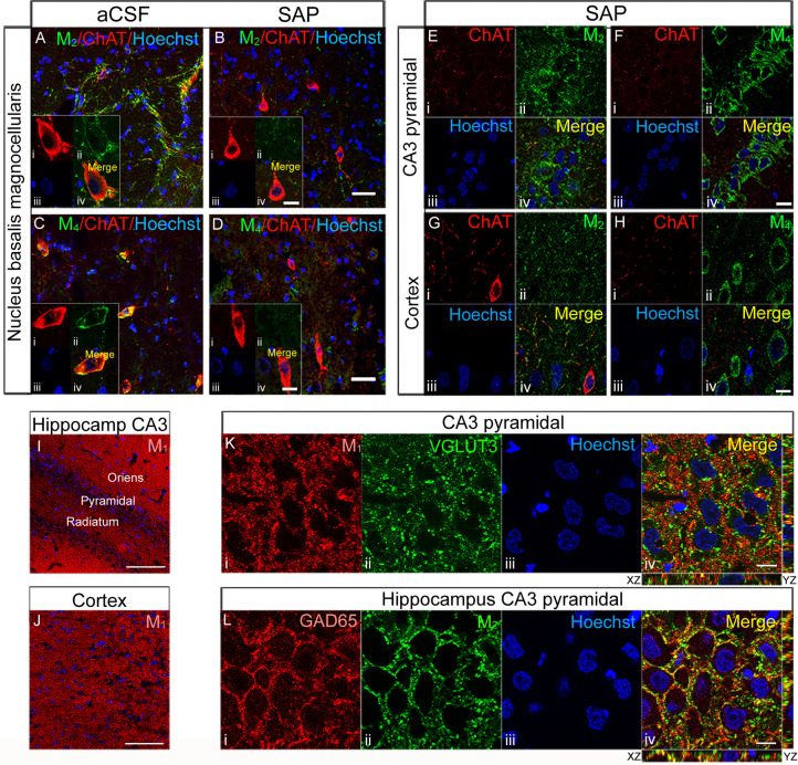
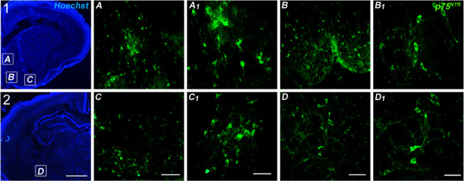
Alzheimer's disease (AD) represents the most common cause of dementia worldwide and has been consistently associated with the loss of basal forebrain cholinergic neurons (BFCNs) leading to impaired cholinergic neurotransmission, aberrant synaptic function, and altered structural lipid metabolism. In this sense, membrane phospholipids (PLs) can be used for de novo synthesis of choline (Ch) for the further obtaining of acetylcholine (ACh) when its availability is compromised. Specific lipid species involved in the metabolism of Ch have been identified as possible biomarkers of phenoconversion to AD. Using a rat model of BFCN lesion, we have evaluated the lipid composition and muscarinic signaling in brain areas related to cognitive processes. The loss of BFCN resulted in alterations of varied lipid species related to Ch metabolism at nucleus basalis magnocellularis (NMB) and cortical projection areas. The activity of muscarinic receptors (mAChR) was decreased in the NMB and increased in the hippocampus according to the subcellular distribution of M1/M2 mAChR which could explain the learning and memory impairment reported in this AD rat model. These results suggest that the modulation of specific lipid metabolic routes could represent an alternative therapeutic strategy to potentiate cholinergic neurotransmission and preserve cell membrane integrity in AD.

Keywords: 192IgG-saporin; Cholinergic; MALDI; autoradiography; lipid; muscarinic receptor.


Conflict of interest statement

The authors declare no competing financial interest.

Figures

Figure 1
Figure 1
(A) Acquisition latency times in the learning trial of the passive avoidance test of SHAM (n = 6), aCSF (n = 36), and SAP (n = 36) rats. (B) Electrical intensity (mA) required to provoke the first audible vocalization when delivering increasing intensities of foot-shock to aCSF (n = 8) and SAP (n = 11) rats (p = ns aCSF vs SAP). (C) Step-through latency times in the retention trial, represented as Kaplan–Meier survival curves. The median step-through latency or the time spent to enter the dark compartment by 50% of the immunotoxin-treated rats was calculated in 102.9 s (aCSF vs SHAM, p = ns; SAP vs aCSF p < 0.001, log-rank test). (D, E) AChE staining in brain sections at three different levels of aCSF and SAP-treated rats. (F) Microphotographs from somatosensory cortex of aCSF and SAP-treated rats at 400-fold magnification, revealing decreased AChE positive fibers in the immunotoxin-treated rats (scale bar = 20 μm). (G) Optical density of AChE expressed as percentages of the striatal levels (used as control area) in aCSF (n = 13) and SAP (n = 13) treated rats: *p < 0.05, **p < 0.01, and ***p < 0.001 SAP vs aCSF-treated rats. Outliers are indicated by black dots or circles in the G panel. MS: medial septum. Cg: cingulate cortex. Mot: motor cortex. Ss: somatosensory cortex. Pir: piriform cortex. Ent: entorhinal cortex. Amyg: amygdaloid nuclei (Bla, basolateral; Ant, anterior; Lat, lateral; Cent, central; Med, medial). DG: dentate gyrus. CA1-CA2-CA3: hippocampal CA1-CA2-CA3 regions. Str: striatum.
Figure 2
Figure 2
Matrix-assisted laser desorption ionization imaging mass spectrometry (MALDI-IMS) of different lipids in brain slices containing the NBM and cortical projections from aCSF (n = 8) and SAP-treated rats (n = 8) which show marked differences in the distribution of certain lipid species following the cholinergic lesion. The intensities were measured in the areas marked with black squares of the somatosensory cortex and the NBM. Scale bar = 5 mm.
Figure 3
Figure 3
Relative abundance of different lipid species in NBM (A) and cortex (B), expressed as percentages of the control group values (aCSF): *p < 0.05, **p < 0.01, and ***p < 0.001, SAP vs aCSF-treated rats.
Figure 4
Figure 4
Hoechst staining (blue) and Iba1 (red), showing coronal representative sections of rat brains in the proximity of the injection site: (A, B, C, D, E) aCSF; (A′, B′, C′, D′, E′) SAP-treated. The inset squares indicate the brain areas where the microglia density is shown: (B, B′) cortical area near the injection; (C, C′) correspondence to the injection site at the NBM nucleus; (D, D′) cingulate cortex; (E, E′) area of thalamic nuclei. Note the higher density of Iba-1 positive cells (microglia) in lesioned animal, which is more noticeable and extended at the lesion site in the NBM. Bar in (A) is 500 μm. Bar in (B) is 100 μm.
Figure 5
Figure 5
[35S]GTPγS autoradiography in rat brain coronal sections at two different levels from Bregma, (A) including NBM and (B) including dorsal hippocampus, obtained from aCSF, left (n = 9), and SAP-treated rats, right (n = 11), that show representative autoradiograms of [35S]GTPγS binding evoked by carbachol (100 μM). This assay is specific to Gi/o coupled receptors; therefore we are measuring the activity mediated by M2/M4 mAChR. The graphs show the mean ± SEM of each group in the different analyzed areas. NBM: nucleus basalis magnocellularis. D gyrus (granular): granular dentate gyrus. CA3: CA3 region of hippocampus. S sensory: somatosensory cortex. [14C]-Microscales were used as standards in nCi/g t.e. Scale bar: 5 mm.
Figure 6
Figure 6
[3H]-Pirenzepine binding (A) and [3H]-oxotremorine binding (B) in rat brain coronal sections obtained from aCSF (n = 7) and SAP-treated rats (n = 9) that show the specific distribution of M1 mAChR and M2 mAChR, respectively. Note the increase of both the M1 mAChR density in the hippocampus (A) and the M2 mAChR density in the deepest layers of cortex (B) and in the anterior amygdala of SAP-treated rats. Note also the loss of both M1 mAChR in the anterior amygdala (A) and M2 mAChR in the NBM (B) following the lesion. The histograms show the mean ± SEM of each group in the different areas analyzed. Scale bar: 5 mm.
Figure 7
Figure 7
Double labeling in sections containing NBM from aCSF (A, C) and 192IgG-saporin-treated (B, D) rats, stained for ChAT (red) and M2 mAChR and M4 mAChR (green) at 200-fold magnification. 192IgG-saporin induced a reduction in BFCN density and in M2/M4 mAChR-immunoreactivity. Scale bars (B, D) = 40 μm. High magnification images show M2 mAChR immunoreactivity surrounding the perikarya (Aii) of the large BFCN (Ai) with a modest degree of colocalization (Aiv), whereas M4 mAChR-immunolabeling, surrounding the perikarya (Cii) of the large BFCN (Ci), shows a high degree of colocalization (Civ). Interestingly, 192IgG-saporin-treated rats show the presence of shrunk ChAT-immunoreactive neurons (Bi, Di), where the loss of M2 and M4 mAChR immunoreactivity is evident (Bii and Dii, respectively). Scale bars (Biv, Div) = 10 μm. Double labeling of consecutive sections containing hippocampal CA3 pyramidal region (E, F) and somatosensory cortex (G, H) from one SAP-treated rat, stained for ChAT (red), M2 mAChR (E and G in green), and M4 mAChR (F and H in green) at 630-fold magnification. The images show a presynaptic distribution of M2 mAChR, delineating the perikarya of the large CA3 pyramidal neurons in basket-like formations (Eii) and the somatodendritic distribution of M4 mAChR in the perikarya of the same neurons (Fii). In the cortex, both M2 (G) and M4 (H) mAChR distribution displays a similar pattern to that observed in CA3. Scale bars (Fiv, Hiv) = 10 μm. M1 mAChR immunolabeling in hippocampal CA3 region (I) and somatosensory cortex (J) from one SAP-treated rat at 200-fold magnification. Scale bars (I, J) = 100 μm. 630-fold magnification images show M1 mAChR (Ki) immunoreactivity distributed surrounding the nuclei (Kiii) in the perikarya of pyramidal neurons with a modest degree of colocalization (Kiv) with the glutamatergic marker VGLUT3 (Kii). M2 mAChRs (Lii) are distributed in presynaptic GABAergic terminals (Li) in basket-like formations surrounding the somatodendritic compartment of pyramidal neurons of CA3 with a high degree of colocalization (Liv). Scale bars = 10 μm.
Figure 8
Figure 8
Low magnification (2.5×) photomicrographs of Hoechst staining in 10 μm tissue slices from P7 rats including the medial septum (MS) (1A), both vertical diagonal band of Broca (1B) and horizontal diagonal band of Broca (1C) and the NBM (2D) (scale bar = 1 mm). P75NTR immunofluorescence in the MS (A, A1). Vertical diagonal band (B, B1). Horizontal diagonal band (C, C1). NBM (D, D1). (C, D) Scale bar = 100 μm. (C1, D1) Scale bar = 50 μm.
Figure 9
Figure 9
Correlation analyses between PC 36:4, PC 38:6, and PC 38:5 with LPC 16:0 and 18:0 in organotypic cultures following three different experimental treatments (vehicle, carbachol 1 μM, and carbachol 1 μM + scopolamine 1 μM): *p < 0.05, **p < 0.01, and ***p < 0.001.

References

    1. Davies P.; Maloney A. J. (1976) Selective Loss of Central Cholinergic Neurons in Alzheimer’s Disease. Lancet 308, 1403.10.1016/S0140-6736(76)91936-X. - DOI - PubMed
    1. Perry E. K.; Gibson P. H.; Blessed G.; Perry R. H.; Tomlinson B. E. (1977) Neurotransmitter Enzyme Abnormalities in Senile Dementia. Choline Acetyltransferase and Glutamic Acid Decarboxylase Activities in Necropsy Brain Tissue. J. Neurol. Sci. 34 (2), 247–265. 10.1016/0022-510X(77)90073-9. - DOI - PubMed
    1. Whitehouse P. J.; Price D. L.; Struble R. G.; Clark A. W.; Coyle J. T.; Delon M. R. (1982) Alzheimer’s Disease and Senile Dementia: Loss of Neurons in the Basal Forebrain. Science 215 (4537), 1237–1239. 10.1126/science.7058341. - DOI - PubMed
    1. Rodríguez-Puertas R.; Pazos A.; Zarranz J. J.; Pascual J. (1994) Selective Cortical Decrease of High-Affinity Choline Uptake Carrier in Alzheimer’s Disease: An Autoradiographic Study Using 3H-Hemicholinium-3. J. Neural Transm.: Parkinson's Dis. Dementia Sect. 8 (3), 161–169. 10.1007/BF02260937. - DOI - PubMed
    1. Rodríguez-Puertas R.; Pascual J.; Vilaró T.; Pazos A. (1997) Autoradiographic Distribution of M1, M2, M3, and M4Muscarinic Receptor Subtypes in Alzheimer’s Disease. Synapse 26 (4), 341–350. 10.1002/(SICI)1098-2396(199708)26:4<341::AID-SYN2>3.0.CO;2-6. - DOI - PubMed

Publication types