. 2021 Aug 1;162(8):bqab103.
doi: 10.1210/endocr/bqab103.

Neuroendocrine Basis for Disrupted Ovarian Cyclicity in Female Mice During Chronic Undernutrition

Affiliations

Neuroendocrine Basis for Disrupted Ovarian Cyclicity in Female Mice During Chronic Undernutrition

Michael J Kreisman et al. Endocrinology. .

Abstract

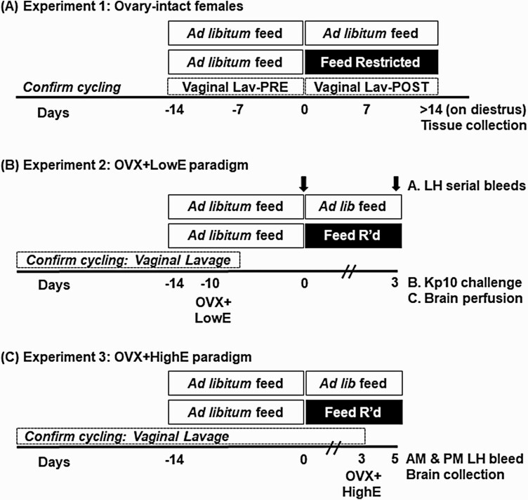
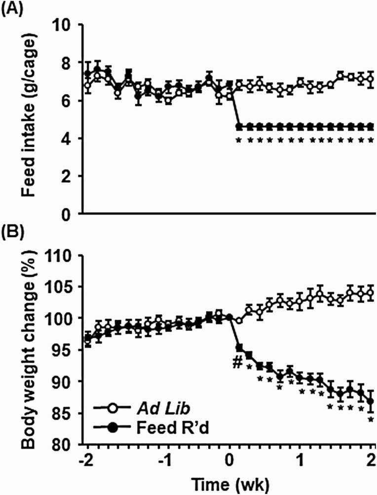
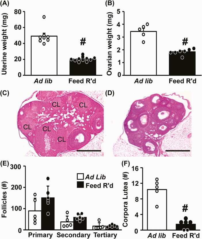
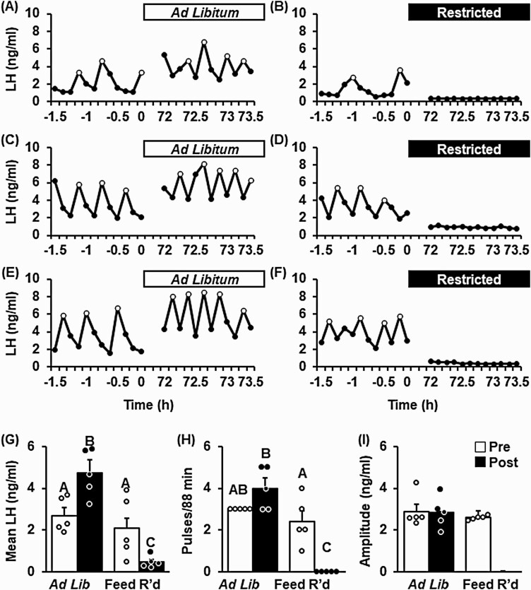
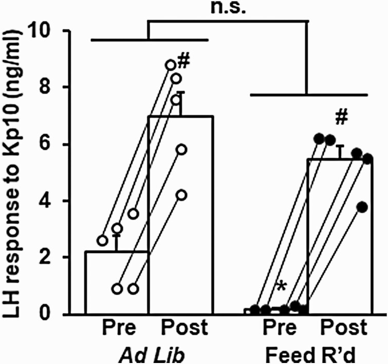
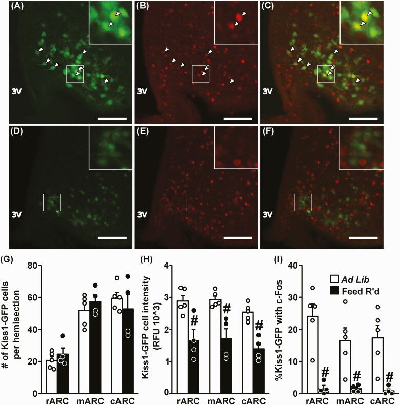
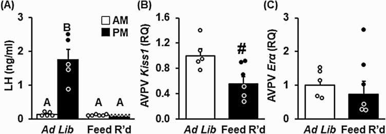
Chronic undernutrition is a type of metabolic stress that impairs reproduction in multiple species. Although energy balance and female reproductive capacity is recognized as tightly coupled, the neuroendocrine loci and molecular mechanisms that mediate ovarian cycle dysfunction during chronic undernutrition in adult females remain poorly understood. Here, we present a series of studies in which we tested the hypothesis that inhibition of kisspeptin (Kiss1) neurons, which are critical for controlling luteinizing hormone (LH) pulses and the preovulatory LH surge in females, underlies the impairment of the ovarian cycle by undernutrition. We first investigated the effect of chronic undernutrition (70% of unrestricted feed intake) on estrous cyclicity in intact female c57bl6 mice. Undernutrition caused a rapid cessation of ovarian cyclicity during the 2-week treatment, suppressing ovarian steroidogenesis and inhibiting ovulation. Using 2 well-defined estradiol-replacement paradigms, we directly tested the hypothesis that undernutrition inhibits Kiss1 neurons in the arcuate nucleus (ARCKiss1), which are required for LH pulses and in the anteroventral periventricular nucleus (AVPVKiss1), which are necessary for LH surge secretion. Undernutrition prevented LH pulses and impaired ARCKiss1 neuronal activation, using c-Fos as a marker, in ovariectomized females subcutaneously implanted with a pellet containing a diestrus-like level of estradiol. In addition, undernutrition completely blocked the estradiol-induced LH surge and diminished Kiss1 messenger RNA abundance, without decreasing estradiol receptor α (Erα), in micropunches of the AVPV. Collectively, these studies demonstrate that undernutrition disrupts ovarian cyclicity in females via impairment both of ARCKiss1 control of LH pulses and AVPVKiss1 induction of the LH surge.

Keywords: feed restriction; gonadotropin-releasing hormone; kisspeptin; luteinizing hormone; metabolism; stress.


Figures

Figure 1.
Figure 1.
Experimental details. Time is depicted as days relative to the initiation of feed restriction or ad libitum feeding in controls, indicated by the horizontal bars. A, In experiment 1, vaginal lavage (Lav) was performed to assess estrous cyclicity in intact females receiving ad libitum feed or feed restriction; animals were euthanized for tissue collection the first day of diestrus that occurred after 14 days of feed restriction or ad libitum (control) feeding. B, Experiment 2 consisted of 3 subexperiments in which females were ovariectomized (OVX) and received a low, diestrus-like, estradiol implant (OVX + LowE) 10 days prior to receiving ad libitum feed or feed restriction. In experiment 2A, frequent blood samples were collected prior to (–1.5 to 0 hours) or following (72-73.5 hours) the onset of treatment, as indicated by the arrows. (Experiment 2B) Blood to evaluate Kp10-induced luteinizing hormone (LH) secretion or (experiment 2C) fixed neural tissue were collected 3 days following onset of ad libitum feed or feed restriction. C, In experiment 3, females were OVX and received a high, LH surge-inducing, estradiol implant (OVX + HighE) and blood and tissues were collected 2 days later.
Figure 2.
Figure 2.
Chronic undernutrition reduces body weight of intact female mice. A, Mean (± SEM) feed intake per female pair depicted as grams/cage, and B, average individual body weight depicted as the percentage change relative to time 0, in females that received control (ad libitum) feed or feed restriction (70% of ad libitum feed intake). Values (mean ± SEM) were analyzed by 2-way analysis of variance with time and treatment as factors; n = 8-11 animals/group. #Significance compared to day 0 in feed-restricted animals (P < .05). *Significance compared to day 0 in feed-restricted animals, as well as significance compared to control animals the same day (P < .05).
Figure 3.
Figure 3.
Chronic undernutrition impairs estrous cyclicity in intact female mice. Representative profiles depicting estrous cyclicity in intact female mice, as measured by vaginal cytology, prior to and during, A and C, ad libitum feeding, or B and D, feed restriction (Restricted/Fd R’d). Number of estrous cycles, E, defined by an estrus-diestrus-estrus transition, and F, average estrous cycle length prior to and during ad libitum feeding or feed restriction. G, Average time spent in each stage of the cycle prior to and during treatment. Values (mean ± SEM) were analyzed by 2-way analysis of variance with time and treatment as factors; n = 8-11 animals/group. Unique letters signify significant differences between values (P < .05). #Time × treatment interaction (P < .05). E, estrus, P, proestrus, D, diestrus, M, metestrus.
Figure 4.
Figure 4.
Diminished ovarian function and anovulation induced by chronic undernutrition. Mean (± SEM) A, uterine weight, and B, ovarian weight collected from diestrus females following 2 weeks of ad libitum feeding or feed restriction (Feed R’d); n = 8-11 (uterine weight) or n = 5-7 (ovarian weight) animals per group. Representative photomicrographs of hematoxylin and eosin–stained fixed ovarian tissue collected from females following C, ad libitum feeding, or D, feed restriction. Scale bar equals 500 μm; CL, corpora lutea. Values (mean ± SEM) for E, primary, secondary, and tertiary follicles, and F, corpora lutea, were quantified in each ovary and analyzed by one-way analysis of variance; n = 5-7 animals/group. #Effect of treatment (P < .05).
Figure 5.
Figure 5.
Undernutrition disrupts luteinizing hormone (LH) pulses in female mice ovariectomized and implanted with a, diestrus-like, estradiol-implant (OVX + LowE). Pattern of pulsatile LH secretion, measured in serial tail-tip blood samples, in 3 representative OVX + LowE mice prior to or following 3 days of A, C, and E, ad libitum feed, or B, D, and F, feed restriction (Feed R’d). LH pulses are identified by open circles. Values (mean ± SEM) for G, mean LH; H, pulse frequency; and I, pulse amplitude were calculated across time (pre [–1.5 to 0 hours], white bars vs post [72-73.5 hours], black bars) and between treatment groups (n = 5 mice/group) by 2-way analysis of variance with time and treatment as factors. Unique letters signify significant differences between values (P < .05). Note, pulse amplitude was not calculated following feed restriction because no pulses were identified during the post period.
Figure 6.
Figure 6.
Undernutrition does not alter the luteinizing hormone (LH) response to exogenous kisspeptin (Kp10). Mean (± SEM) LH measured 10 minutes prior to and following Kp10 (2 µg/g, intraperitoneally) administered to female mice ovariectomized and implanted with a, diestrus-like, estradiol-implant (OVX + LowE) following 3 days of ad libitum feeding (white circles) or feed restriction (black circles). Lines connect the pre and post sample for each animal. *Effect of treatment (P < .05). #Effect of time (Pre/Post) (P < .05). No significant treatment × time interaction was identified (P > .05).
Figure 7.
Figure 7.
Undernutrition reduces Kiss1 expression and neuronal activation in the arcuate nucleus. Representative photomicrographs depicting staining for A and D, Kiss1 (green); B and E, c-Fos (red); and C and F, dual-labeled Kiss1/c-Fos cells in the middle ARC of OVX + LowE Kiss1hrGFP mice following A to C, ad libitum feeding, or D to F, 3 days of feed restriction (Feed R’d). White box indicates location of zoomed panels. White arrowheads indicate dual-labeled Kiss1/c-Fos cells. Scale bar equals 100 μm. Brightness and contrast were adjusted similarly in images from control and feed-restricted animals. G, Number of Kiss1 cells per hemisection; H, Kiss1 cell intensity per section; I, percentage of Kiss1 cells with c-Fos per hemisection following ad libitum feeding (white bars) or feed restriction (black bars). Regions of the ARC are referred to as rostral (r), middle (m), and caudal (c); 3V, third ventricle. Values (mean ± SEM) were analyzed by one-way analysis of variance. #Effect of treatment (P < .05).
Figure 8.
Figure 8.
Undernutrition lowers Kiss1 neurons in the anteroventral periventricular nucleus (AVPVKiss1) expression and prevents the estradiol-induced luteinizing hormone (LH) surge. A, Blood was collected in the morning (am, 1000 hours) and evening (pm, 1800 hours) of the expected LH surge in female mice following 5 days of ad libitum feeding or feed restriction (Feed R’d) and analyzed by 2-way analysis of variance (ANOVA) with time and treatment as factors. Unique letters signify significant differences between values (P < .05). Kiss1 and estradiol receptor α (Erα) gene expression in AVPV micropunches, collected following ad libitum feeding (white bars) or feed restriction (black bars), were analyzed by 1-way ANOVA. #Effect of treatment (P < .05); n = 5-6/group. Abbreviation: RQ, Relative Quantity.

References

    1. Knorr D, Khoo CH. COVID-19 and food: challenges and research needs. Front Nutr. 2020;7:598913. - PMC - PubMed
    1. Sun J, Shen X, Liu H, Lu S, Peng J, Kuang H. Caloric restriction in female reproduction: is it beneficial or detrimental? Reprod Biol Endocrinol. 2021;19(1):1. - PMC - PubMed
    1. Iwasa T, Matsuzaki T, Yano K, et al. Effects of low energy availability on reproductive functions and their underlying neuroendocrine mechanisms. J Clin Med. 2018;7(7):166. - PMC - PubMed
    1. Bronson FH, Marsteller FA. Effect of short-term food deprivation on reproduction in female mice. Biol Reprod. 1985;33(3):660-667. - PubMed
    1. Foster DL, Ebling FJ, Micka AF, et al. Metabolic interfaces between growth and reproduction. I. Nutritional modulation of gonadotropin, prolactin, and growth hormone secretion in the growth-limited female lamb. Endocrinology. 1989;125(1):342-350. - PubMed

Publication types

Substances