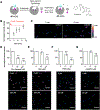
Visualizing the Dynamic Metalation State of New Delhi Metallo-β-lactamase-1 in Bacteria Using a Reversible Fluorescent Probe
- PMID: 34038127
- PMCID: PMC8230704
- DOI: 10.1021/jacs.1c00290
Visualizing the Dynamic Metalation State of New Delhi Metallo-β-lactamase-1 in Bacteria Using a Reversible Fluorescent Probe
Abstract
New Delhi metallo-β-lactamase (NDM) grants resistance to a broad spectrum of β-lactam antibiotics, including last-resort carbapenems, and is emerging as a global antibiotic resistance threat. Limited zinc availability adversely impacts the ability of NDM-1 to provide resistance, but a number of clinical variants have emerged that are more resistant to zinc scarcity (e.g., NDM-15). To provide a novel tool to better study metal ion sequestration in host-pathogen interactions, we describe the development of a fluorescent probe that reports on the dynamic metalation state of NDM within Escherichia coli. The thiol-containing probe selectively coordinates the dizinc metal cluster of NDM and results in a 17-fold increase in fluorescence intensity. Reversible binding enables competition and time-dependent studies that reveal fluorescence changes used to detect enzyme localization, substrate and inhibitor engagement, and changes to metalation state through the imaging of live E. coli using confocal microscopy. NDM-1 is shown to be susceptible to demetalation by intracellular and extracellular metal chelators in a live-cell model of zinc dyshomeostasis, whereas the NDM-15 metalation state is shown to be more resistant to zinc flux. The development of this reversible turn-on fluorescent probe for the metalation state of NDM provides a new tool for monitoring the impact of metal ion sequestration by host defense mechanisms and for detecting inhibitor-target engagement during the development of therapeutics to counter this resistance determinant.
Figures


Similar articles
-
Benzimidazole and Benzoxazole Zinc Chelators as Inhibitors of Metallo-β-Lactamase NDM-1.ChemMedChem. 2021 Feb 17;16(4):654-661. doi: 10.1002/cmdc.202000607. Epub 2020 Nov 19. ChemMedChem. 2021. PMID: 33211374 Free PMC article.
-
Characterization of purified New Delhi metallo-β-lactamase-1.Biochemistry. 2011 Nov 22;50(46):10102-13. doi: 10.1021/bi201449r. Epub 2011 Nov 1. Biochemistry. 2011. PMID: 22029287
-
Evolution of New Delhi metallo-β-lactamase (NDM) in the clinic: Effects of NDM mutations on stability, zinc affinity, and mono-zinc activity.J Biol Chem. 2018 Aug 10;293(32):12606-12618. doi: 10.1074/jbc.RA118.003835. Epub 2018 Jun 16. J Biol Chem. 2018. PMID: 29909397 Free PMC article.
-
Enzyme Inhibitors: The Best Strategy to Tackle Superbug NDM-1 and Its Variants.Int J Mol Sci. 2021 Dec 24;23(1):197. doi: 10.3390/ijms23010197. Int J Mol Sci. 2021. PMID: 35008622 Free PMC article. Review.
-
Recent research and development of NDM-1 inhibitors.Eur J Med Chem. 2021 Nov 5;223:113667. doi: 10.1016/j.ejmech.2021.113667. Epub 2021 Jun 24. Eur J Med Chem. 2021. PMID: 34225181 Review.
Cited by
-
Exploring antibiotic resistance with chemical tools.Chem Commun (Camb). 2023 May 18;59(41):6148-6158. doi: 10.1039/d3cc00759f. Chem Commun (Camb). 2023. PMID: 37039397 Free PMC article. Review.
-
Proteomic strategies to interrogate the Fe-S proteome.Biochim Biophys Acta Mol Cell Res. 2024 Oct;1871(7):119791. doi: 10.1016/j.bbamcr.2024.119791. Epub 2024 Jun 25. Biochim Biophys Acta Mol Cell Res. 2024. PMID: 38925478 Review.
-
Probing metalloenzyme dynamics in living systems: Contemporary advances in fluorescence imaging tools and applications.Curr Opin Chem Biol. 2024 Aug;81:102475. doi: 10.1016/j.cbpa.2024.102475. Epub 2024 Jun 8. Curr Opin Chem Biol. 2024. PMID: 38852500 Free PMC article. Review.
-
High-level nitrofurantoin resistance in a clinical isolate of Klebsiella pneumoniae: a comparative genomics and metabolomics analysis.mSystems. 2024 Jan 23;9(1):e0097223. doi: 10.1128/msystems.00972-23. Epub 2023 Dec 11. mSystems. 2024. PMID: 38078757 Free PMC article.
-
Binding of Dual-Function Hybridized Metal-Organic Capsules to Enzymes for Cascade Catalysis.JACS Au. 2022 Jul 6;2(7):1736-1746. doi: 10.1021/jacsau.2c00322. eCollection 2022 Jul 25. JACS Au. 2022. PMID: 35911460 Free PMC article.
References
-
- Linciano P; Centron L; Gianquinto E; Spyrakis F; Tondi D Ten Years with New Delhi Metallo-β-Lactamase-1 (NDM-1): From Structural Insights to Inhibitor Design. ACS Infect. Dis 2019, 5, 9. - PubMed
-
- Thomas PW; et al. Characterization of Purified New Delhi Metallo-β-lactamase-1. Biochemistry 2011, 50, 10102. - PubMed

