. 2021 May;49(5):3000605211013179.
doi: 10.1177/03000605211013179.

Susceptibility-diffusion mismatch correlated with leptomeningeal collateralization in large vessel occlusion stroke

Affiliations

Susceptibility-diffusion mismatch correlated with leptomeningeal collateralization in large vessel occlusion stroke

Haifei Jiang et al. J Int Med Res. 2021 May.

Abstract

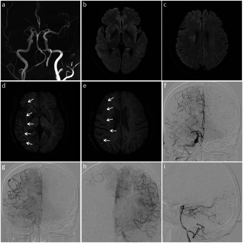
Objective: To investigate the relationship between asymmetric prominent hypointense vessels (prominent vessel sign, PVS) on susceptibility-weighted imaging (SWI) and leptomeningeal collateralization in patients with acute ischemic stroke due to large vessel occlusion.

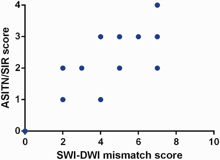
Methods: We retrospectively enrolled patients with M1 segment occlusion of the middle cerebral artery who underwent emergency magnetic resonance imaging and digital subtraction angiography within 24 hours from stroke onset. The extent of PVS on SWI was assessed using the Alberta Stroke Program Early CT Score (ASPECTS). Leptomeningeal collateralization on digital subtraction angiography images was assessed using the American Society of Interventional and Therapeutic Neuroradiology/Society of Interventional Radiology (ASITN/SIR) scale. Spearman's rank correlation test was performed to explore the correlation of ASITN/SIR scores with SWI-ASPECTS and SWI-diffusion-weighted imaging (DWI) mismatch scores.

Results: Thirty-five patients were enrolled. There was no significant correlation between SWI-ASPECTS and ASITN/SIR scores. However, SWI-DWI mismatch scores were positively correlated with ASITN/SIR scores.

Conclusion: The range of PVS on SWI did not closely reflect the collateral status, while the range of SWI-DWI mismatch was significantly correlated with the leptomeningeal collateralization. In patients with acute anterior circulation stroke due to large vessel occlusion, larger SWI-DWI mismatch was associated with better leptomeningeal collaterals.

Keywords: Acute ischemic stroke; diffusion-weighted imaging; large vessel occlusion; leptomeningeal collateralization; prominent vessel sign; susceptibility-weighted imaging.


Conflict of interest statement

Declaration of conflicting interest: The authors declare that there is no conflict of interest.

Figures

Figure 1.
Figure 1.
Images from a patient with a low SWI-DWI mismatch score and poor leptomeningeal collateralization. (a) MRA showed occlusion of the right internal carotid and middle cerebral arteries. (b, c) DWI revealed restricted diffusion in the internal capsule, insular cortex, and M1 to M6 zones of the right middle cerebral artery territory. (d, e) SWI showed the prominent vessel sign (white arrows) in seven regions (right insular and M1 to M6). The SWI-ASPECTS value was 3 (10 − 7 = 3), and the SWI-DWI mismatch score was 0 (7 − 7 = 0). (f) DSA showed occlusion of the right terminal internal carotid artery. (g) Microcatheterography confirmed a T-shaped occlusion at the end of the internal carotid artery (including the proximal M1 of the middle cerebral artery). (f–i) There was no collateral blood supply to the right middle cerebral artery supplying area, and the ASITN/SIR score was 0. SWI, susceptibility-weighted imaging; DWI, diffusion-weighted imaging; MRA, magnetic resonance angiography; ASPECTS, Alberta Stroke Program Early CT Score; DSA, digital subtraction angiography; ASITN/SIR, American Society of Interventional and Therapeutic Neuroradiology/Society of Interventional Radiology.
Figure 2.
Figure 2.
Images from a patient with a high SWI-DWI mismatch score and good leptomeningeal collateralization. (a) MRA showed M1 segment occlusion of the right middle cerebral artery. (b, c) DWI revealed restricted diffusion in the right lentiform nucleus and paraventricular region. (d, e) SWI showed the prominent vessel sign (white arrows) in seven regions (right insular and M1 to M6). The SWI-ASPECTS value was 3 (10 − 7 = 3), and the SWI-DWI mismatch score was 7 (7 − 0 = 7). (f) DSA indicated occlusion of the proximal M1 segment of the right middle cerebral artery. (f–i) The ischemic area was compensated by the leptomeningeal collaterals of the right anterior cerebral artery, and the ASITN/SIR score was 3. SWI, susceptibility-weighted imaging; DWI, diffusion-weighted imaging; MRA, magnetic resonance angiography; ASPECTS, Alberta Stroke Program Early CT Score; DSA, digital subtraction angiography; ASITN/SIR, American Society of Interventional and Therapeutic Neuroradiology/Society of Interventional Radiology.
Figure 3.
Figure 3.
Scatter plot showing the correlation between the SWI-DWI mismatch score and the ASITN/SIR score. The SWI-DWI mismatch score was positively correlated with the ASITN/SIR score (Spearman test, r = 0.818, P < 0.001). SWI, susceptibility-weighted imaging; DWI, diffusion-weighted imaging; ASITN/SIR, American Society of Interventional and Therapeutic Neuroradiology/Society of Interventional Radiology.

Similar articles

Cited by

References

    1. Liebeskind DS. Collateral circulation. Stroke 2003; 34: 2279–2284. - PubMed
    1. Bang OY, Goyal M, Liebeskind DS. Collateral circulation in ischemic stroke: assessment tools and therapeutic strategies. Stroke 2015; 46: 3302–3309. - PMC - PubMed
    1. Ginsberg MD. The cerebral collateral circulation: relevance to pathophysiology and treatment of stroke. Neuropharmacology 2018; 134: 280–292. - PubMed
    1. Haacke EM, Xu Y, Cheng YC, et al.. Susceptibility weighted imaging (SWI). Magn Reson Med 2004; 52: 612–618. - PubMed
    1. Chen CY, Chen CI, Tsai FY, et al.. Prominent vessel sign on susceptibility-weighted imaging in acute stroke: prediction of infarct growth and clinical outcome. PLoS One 2015; 10: e0131118. - PMC - PubMed