

4S-DT: Self-Supervised Super Sample Decomposition for Transfer Learning With Application to COVID-19 Detection
- PMID: 34038371
- PMCID: PMC8544943
- DOI: 10.1109/TNNLS.2021.3082015
4S-DT: Self-Supervised Super Sample Decomposition for Transfer Learning With Application to COVID-19 Detection
Abstract
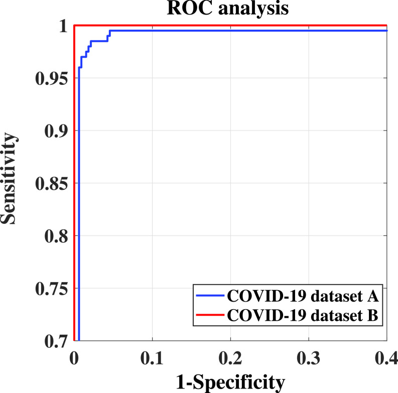
Due to the high availability of large-scale annotated image datasets, knowledge transfer from pretrained models showed outstanding performance in medical image classification. However, building a robust image classification model for datasets with data irregularity or imbalanced classes can be a very challenging task, especially in the medical imaging domain. In this article, we propose a novel deep convolutional neural network, which we called self-supervised super sample decomposition for transfer learning (4S-DT) model. The 4S-DT encourages a coarse-to-fine transfer learning from large-scale image recognition tasks to a specific chest X-ray image classification task using a generic self-supervised sample decomposition approach. Our main contribution is a novel self-supervised learning mechanism guided by a super sample decomposition of unlabeled chest X-ray images. 4S-DT helps in improving the robustness of knowledge transformation via a downstream learning strategy with a class-decomposition (CD) layer to simplify the local structure of the data. The 4S-DT can deal with any irregularities in the image dataset by investigating its class boundaries using a downstream CD mechanism. We used 50000 unlabeled chest X-ray images to achieve our coarse-to-fine transfer learning with an application to COVID-19 detection, as an exemplar. The 4S-DT has achieved a high accuracy of 99.8% on the larger of the two datasets used in the experimental study and an accuracy of 97.54% on the smaller dataset, which was enriched by augmented images, out of which all real COVID-19 cases were detected.
Figures


References
-
- Dandil E., Cakiroglu M., Eksi Z., Ozkan M., Kurt O. K., and Canan A., “Artificial neural network-based classification system for lung nodules on computed tomography scans,” in Proc. 6th Int. Conf. Soft Comput. Pattern Recognit. (SoCPaR), Aug. 2014, pp. 382–386.
-
- Kuruvilla J. and Gunavathi K., “Lung cancer classification using neural networks for CT images,” Comput. Methods Programs Biomed., vol. 113, no. 1, pp. 202–209, Jan. 2014. - PubMed
-
- Manikandan T. and Bharathi N., “Lung cancer detection using fuzzy auto-seed cluster means morphological segmentation and SVM classifier,” J. Med. Syst., vol. 40, no. 7, p. 181, Jul. 2016. - PubMed
-
- Sangamithraa P. B. and Govindaraju S., “Lung tumour detection and classification using EK-mean clustering,” in Proc. Int. Conf. Wireless Commun., Signal Process. Netw. (WiSPNET), Mar. 2016, pp. 2201–2206.
MeSH terms
LinkOut - more resources
Full Text Sources
Other Literature Sources
Medical
