. 2021 May 26;13(10):13460-13473.
doi: 10.18632/aging.203111. Epub 2021 May 26.

Aging is associated with glial senescence in the brainstem - implications for age-related sympathetic overactivity

Affiliations

Aging is associated with glial senescence in the brainstem - implications for age-related sympathetic overactivity

Priya Balasubramanian et al. Aging (Albany NY). .

Abstract

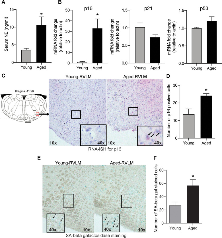
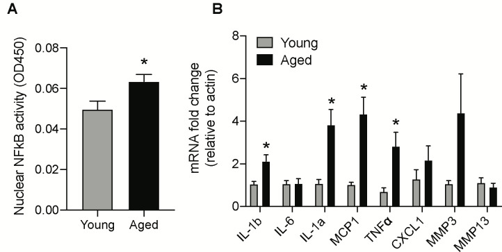
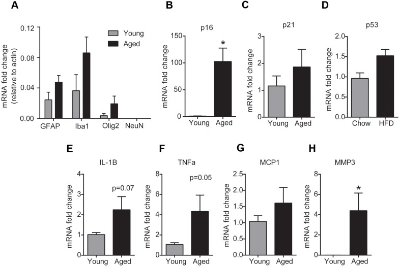
Accumulating evidence suggests that the sympathetic nervous system (SNS) overactivity plays a crucial role in age-related increase in the risk for cardiovascular diseases such as hypertension, myocardial infarction, stroke and heart diseases. Previous studies indicate that neuroinflammation in key brainstem regions that regulate sympathetic outflow plays a pathogenic role in aging-mediated sympathoexcitation. However, the molecular mechanisms underlying this phenomenon are not clear. While senescent cells and their secretory phenotype (SASP) have been implicated in the pathogenesis of several age-related diseases, their role in age-related neuroinflammation in the brainstem and SNS overactivity has not been investigated. To test this, we isolated brainstems from young (2-4 months) and aged (24 months) male C57BL/6J mice and assessed senescence using a combination of RNA-in situ hybridization, PCR analysis, multiplex assay and SA-β gal staining. Our results show significant increases in p16Ink4a expression, increased activity of SA-β gal and increases in SASP levels in the aged brainstem, suggesting age-induced senescence in the brainstem. Further, analysis of senescence markers in glial cells enriched fraction from fresh brainstem samples demonstrated that glial cells are more susceptible to senesce with age in the brainstem. In conclusion, our study suggests that aging induces glial senescence in the brainstem which likely causes inflammation and SNS overactivity.

Keywords: aging; brainstem; glial cells; senescence; sympathetic nervous system.


Conflict of interest statement

CONFLICTS OF INTEREST: The authors declare that they have no conflicts of interest.

Figures

Figure 1
Figure 1
Age-induced changes in serum NE and senescence markers in the brainstem. (A) Changes in serum NE levels in young and aged animals measured using a commercial ELISA kit (mean±SE, n=4-6/group). (B) Real-time PCR analysis of gene expression levels of senescence markers p16, p21 and p53 (n=4/group) (C) Representative images of RNA-ISH showing p16-positive cells in the RVLM of the brainstem in young and aged animals. (D) Semi-quantitative analysis measuring the number of p16-positive cells in the RVLM by RNA-ISH. (E) Representative images of SA-β gal staining in the brainstem and (F) Quantification of cells positive for SA-β gal staining in the RVLM of the brainstem. *denotes a significant difference (p < 0.05) from young animals.
Figure 2
Figure 2
Age-induced changes in NF-κB activity and mRNA levels of SASP factors in the brainstem. (A) NF-κB DNA-binding capacity measured by ELISA in nuclear protein extracted from the brainstem of young and aged animals. (B) Gene expression levels of SASP factors in the brainstem measured by real-time PCR analysis. Data are expressed as mean±SE, n=4-5/group. *denotes a significant difference (p < 0.05) from young animals.
Figure 3
Figure 3
Aging induces glial senescence in the brainstem. (A) Purity of glial cell-enriched fraction assessed by real-time PCR analysis. (BH) Gene expression analysis of senescence markers in the glial cell-enriched fraction from young and aged brainstem. Data are expressed as mean±SE, n=4-5/group. *denotes a significant difference (p < 0.05) from young animals.

References

    1. North BJ, Sinclair DA. The intersection between aging and cardiovascular disease. Circ Res. 2012; 110:1097–108. 10.1161/CIRCRESAHA.111.246876 - DOI - PMC - PubMed
    1. Mozaffarian D, Benjamin EJ, Go AS, Arnett DK, Blaha MJ, Cushman M, Das SR, de Ferranti S, Després JP, Fullerton HJ, Howard VJ, Huffman MD, Isasi CR, et al., and Writing Group Members, American Heart Association Statistics Committee, and Stroke Statistics Subcommittee. Heart Disease and Stroke Statistics-2016 Update: A Report From the American Heart Association. Circulation. 2016; 133:e38–360. 10.1161/CIR.0000000000000350 - DOI - PubMed
    1. Hotta H, Uchida S. Aging of the autonomic nervous system and possible improvements in autonomic activity using somatic afferent stimulation. Geriatr Gerontol Int. 2010. (Suppl 1); 10:S127–36. 10.1111/j.1447-0594.2010.00592.x - DOI - PubMed
    1. Esler M, Hastings J, Lambert G, Kaye D, Jennings G, Seals DR. The influence of aging on the human sympathetic nervous system and brain norepinephrine turnover. Am J Physiol Regul Integr Comp Physiol. 2002; 282:R909–16. 10.1152/ajpregu.00335.2001 - DOI - PubMed
    1. Seals DR, Dinenno FA. Collateral damage: cardiovascular consequences of chronic sympathetic activation with human aging. Am J Physiol Heart Circ Physiol. 2004; 287:H1895–905. 10.1152/ajpheart.00486.2004 - DOI - PubMed

Publication types

LinkOut - more resources