. 2021 May 26;18(5):e1003632.
doi: 10.1371/journal.pmed.1003632. eCollection 2021 May.

Evaluation of splenic accumulation and colocalization of immature reticulocytes and Plasmodium vivax in asymptomatic malaria: A prospective human splenectomy study

Affiliations

Evaluation of splenic accumulation and colocalization of immature reticulocytes and Plasmodium vivax in asymptomatic malaria: A prospective human splenectomy study

Steven Kho et al. PLoS Med. .

Abstract

Background: A very large biomass of intact asexual-stage malaria parasites accumulates in the spleen of asymptomatic human individuals infected with Plasmodium vivax. The mechanisms underlying this intense tropism are not clear. We hypothesised that immature reticulocytes, in which P. vivax develops, may display high densities in the spleen, thereby providing a niche for parasite survival.

Methods and findings: We examined spleen tissue in 22 mostly untreated individuals naturally exposed to P. vivax and Plasmodium falciparum undergoing splenectomy for any clinical indication in malaria-endemic Papua, Indonesia (2015 to 2017). Infection, parasite and immature reticulocyte density, and splenic distribution were analysed by optical microscopy, flow cytometry, and molecular assays. Nine non-endemic control spleens from individuals undergoing spleno-pancreatectomy in France (2017 to 2020) were also examined for reticulocyte densities. There were no exclusion criteria or sample size considerations in both patient cohorts for this demanding approach. In Indonesia, 95.5% (21/22) of splenectomy patients had asymptomatic splenic Plasmodium infection (7 P. vivax, 13 P. falciparum, and 1 mixed infection). Significant splenic accumulation of immature CD71 intermediate- and high-expressing reticulocytes was seen, with concentrations 11 times greater than in peripheral blood. Accordingly, in France, reticulocyte concentrations in the splenic effluent were higher than in peripheral blood. Greater rigidity of reticulocytes in splenic than in peripheral blood, and their higher densities in splenic cords both suggest a mechanical retention process. Asexual-stage P. vivax-infected erythrocytes of all developmental stages accumulated in the spleen, with non-phagocytosed parasite densities 3,590 times (IQR: 2,600 to 4,130) higher than in circulating blood, and median total splenic parasite loads 81 (IQR: 14 to 205) times greater, accounting for 98.7% (IQR: 95.1% to 98.9%) of the estimated total-body P. vivax biomass. More reticulocytes were in contact with sinus lumen endothelial cells in P. vivax- than in P. falciparum-infected spleens. Histological analyses revealed 96% of P. vivax rings/trophozoites and 46% of schizonts colocalised with 92% of immature reticulocytes in the cords and sinus lumens of the red pulp. Larger splenic cohort studies and similar investigations in untreated symptomatic malaria are warranted.

Conclusions: Immature CD71+ reticulocytes and splenic P. vivax-infected erythrocytes of all asexual stages accumulate in the same splenic compartments, suggesting the existence of a cryptic endosplenic lifecycle in chronic P. vivax infection. Findings provide insight into P. vivax-specific adaptions that have evolved to maximise survival and replication in the spleen.


Conflict of interest statement

The authors have declared that no competing interests exist.

Figures

Fig 1
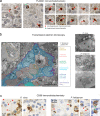
Fig 1. Counting malaria parasites on Giemsa-stained spleen sections of human spleens.
Tissue sections stained with Giemsa were analysed in P. vivax- and P. falciparum-infected spleens at 400× magnification using a Carl Zeiss AxioScan Z1. Representative P. vivax-infected spleen sections are from patient #1 and P. falciparum from patient #17, with staging key of non-phagocytosed P. vivax and P. falciparum shown below these panels (a, asexuals) (a, b). Tissue compartments were categorised into white-pulp non-circulatory spaces and perifollicular zones, and red-pulp sinus lumen and cords. An uninfected control human spleen was perfused with P. falciparum lab strain cultures and had Giemsa-stained sections examined to validate the appearance of asexual-stage Plasmodium (c). Phagocytosed parasites (a–c) and pigmented phagocytes (d) were observed in Giemsa-stained spleen sections from infected individuals with representative images shown for each species.
Fig 2
Fig 2. Confirming the integrity of non-phagocytosed IEs by immunohistochemistry and electron microscopy.
Antibodies against PvAMA1, a marker for P. vivax merozoites/mature stages staining red, were tested on spleen sections from patient #1 (a) and others (S4D Fig) to confirm previous findings [29] on the presence of mature P. vivax stages. Spleen tissue from patients in the cohort was also examined by transmission electron microscopy to identify non-phagocytosed malaria parasites (p) inside erythrocytes (i), with representative images in both P. vivax (patient #2) and P. falciparum (patients #5 and #17) (b). IEs in the spleen were largely localised outside of macrophages (m) as illustrated in P. vivax- (patient #18) and P. falciparum-infected (patient #17) spleen sections stained with macrophage marker CD68, including examples of phagocytosed IEs shown on the right (c). IE, infected erythrocyte; m, macrophages; p, parasites; PvAMA1, Pv apical membrane antigen-1.
Fig 3
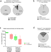
Fig 3. Splenic accumulation and distribution of non-phagocytosed IEs in cohort patients.
Non-phagocytosed parasites in peripheral blood and spleen sections were counted by microscopy using an Olympus CX31 microscope. Stages were categorised into asexual, sexual, and unclassifiable for Pv (n = 6) and Pf (n = 9). In Pv and Pf, all individuals had asexual stages present. Sexual stages were reported in 1 Pv- and 6 Pf-infected individuals, and unclassifiable stages reported in 2 Pv- and 6 Pf-infected individuals, with the median distributions summarised as shown (a). As an additional comparator to the spleen, Pv biomass in the bone marrow was conservatively estimated based on a previous human study comparing relative Pv parasitaemia in the bone marrow to peripheral blood [20] (*). The median percentage of non-phagocytosed asexual Pv biomass found in the spleen, peripheral blood, and bone marrow was calculated (b). The median estimated non-phagocytosed spleen-to-peripheral asexual parasite density and biomass ratios were compared between 6 Pv and 7 Pf (c) (^reported previously [29]). The median distribution of asexual Pv stages in the splenic architecture was categorised into those found in the RP cords and sinus lumens, and WP non-circulatory spaces and perifollicular zones (d). Two individuals had <0.3% of asexual Pv in WP non-circulatory spaces. In panels a and d, the sum of segments in each pie chart may not add up to 100% due to medians in one of the groups being zero despite some individuals having values >0%. In panel c, medians, interquartile ranges, and ranges are shown, compared using the Mann–Whitney test (p-value <0.05 considered significant). IE, infected erythrocyte; Pf, Plasmodium falciparum; Pv, Plasmodium vivax; RP, red-pulp; WP, white-pulp.
Fig 4
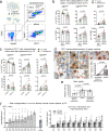
Fig 4. Increased density of immature CD71+ reticulocytes in human spleens.
Samples were magnetically enriched for reticulocytes (retics) and phenotyped by CD71 FC (a). The median retic purity in enriched samples was 95.8% [IQR: 94.3%–98.5%] for PB and 56.8% [IQR: 53.4%–72.6%] for sliced-spleen blood. Numbers as a percentage of CD71+ retics and per 105 or 106 RBCs were determined for CD71 low-, intermediate- and high-expressing retics in PB and the spleen (b) (n = 10 pairs). In addition, CD71+ retic populations in PB several months after splenectomy (median post-splenectomy retic enrichment purity of 84.0% [IQR: 71.9%–86.6%]) was compared to PB at surgery (n = 10 pairs), presented as a percentage of CD71+ retics and as absolute counts per μL blood (calculated based on automated RBC counts that were available for PB) (c). CD71+ retics were visualised on spleen section by CD71 immunohistochemistry (patient #4 section shown) and counted based on guidelines derived from CD71 staining of a retic-enriched blood pellet (d). CD71+ retics concentrations in the spleen by immunohistochemistry was compared to CD71+ retic concentrations in PB determined by FC (n = 10 pairs) (d). Peripheral and splenic retic deformability in 6 patients was determined by microsphiltration and presented as retention rates (e). In France, ex vivo flushing of 9 uninfected control spleens were performed (f). For each of the first 5 spleens, a PB fraction was collected either from the basilic or splenic vein, followed by 5–6 consecutive SW fractions from 350–500 mL of flushing buffer. With initial results indicating a trend towards higher proportions of immature retics in the last SW fractions, the volume of flushing buffer was increased to 500–650 mL for each of the next 4 spleens, resulting in 8–9 SW fractions being collected. Immature CD71+ retics were quantitated in each fraction by FC (see S2D Fig), normalised, and compared to PB (f, left). In the last 4 spleens, retics were phenotyped into CD71 negative, low-, intermediate-, and high-expression (f, right). Paired datapoints are connected by lines. Bars in all panels represent medians. Error bars in f are interquartile ranges. The Wilcoxon test was used for all statistical comparisons (*p < 0.05 considered statistically significant). FC, flow cytometry; PB, peripheral blood; RBC, red blood cell; SW, spleen wash.
Fig 5
Fig 5. Immature CD71+ reticulocytes (retics) and non-phagocytosed Pv-IEs colocalise in specific splenic compartments.
The density and total biomass of CD71+ retics and non-phagocytosed Pv developmental stages (n = 6) were determined in the red-pulp SL, cords, PFZ, and white-pulp NCS (a). Non-phagocytosed Pv parasites were categorised into 3 groups (rings/trophozoites, schizonts, gametocytes), and a fourth group of those with unclassifiable stages. The cumulative distribution of CD71+ retics and Plasmodium developmental stages in the splenic architecture was calculated in individuals in whom intrasplenic parasites were visualised (b, left). The distribution of CD71+ retics in an uninfected spleen is also shown (b, right). In the splenic SL, a large proportion of CD71+ retics were apparently adherent to endothelial cells on the luminal side as observed by CD71 immunohistochemistry (c, left, representative image from Pv patient #4). The number of those apparently adherent (adh) and non-adherent (non-adh) were expressed as a percentage of red-pulp CD71+ retics in Pv as well as Pf, and compared as paired data (connected lines) using the Wilcoxon test (c, middle). The ratio of adherent-to-non-adherent CD71+ retics in the SL was compared between Pv and Pf using the Mann–Whitney test (c, right). A p-value <0.05 was considered significant. Data in a, b (left), and c (right) are individual datapoints with median and interquartile range. adh, adherent; NCS, non-circulatory space; non-adh, non-adherent; Pf, Plasmodium falciparum; PFZ, perifollicular zone; Pv, Plasmodium vivax; RBC, red blood cell; SL, sinus lumen.

Similar articles

Cited by

References

    1. WHO. World Malaria Report 2020. Geneva: World Health Organisation, 2020.
    1. Garrido-Cardenas JA, Cebrián-Carmona J, González-Cerón L, Manzano-Agugliaro F, Mesa-Valle C. Analysis of Global Research on Malaria and Plasmodium vivax. Int J Environ Res Public Health. 2019;16(11):1928. 10.3390/ijerph16111928 . - DOI - PMC - PubMed
    1. Galinski MR, Barnwell JW. Plasmodium vivax: who cares? Malar J. 2008;7(1):S9. 10.1186/1475-2875-7-S1-S9 - DOI - PMC - PubMed
    1. Price RN, Commons RJ, Battle KE, Thriemer K, Mendis K. Plasmodium vivax in the Era of the Shrinking P. falciparum Map. Trends Parasitol. 2020;36(6):560–70. 10.1016/j.pt.2020.03.009 - DOI - PMC - PubMed
    1. Genton B, D’Acremont V, Rare L, Baea K, Reeder JC, Alpers MP, et al.. Plasmodium vivax and Mixed Infections Are Associated with Severe Malaria in Children: A Prospective Cohort Study from Papua New Guinea. PLoS Med. 2008;5(6):e127. 10.1371/journal.pmed.0050127 - DOI - PMC - PubMed

Publication types