. 2021 May 26;16(5):e0250429.
doi: 10.1371/journal.pone.0250429. eCollection 2021.

DSPP dosage affects tooth development and dentin mineralization

Affiliations

DSPP dosage affects tooth development and dentin mineralization

Dandrich Lim et al. PLoS One. .

Abstract

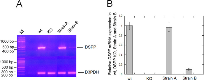
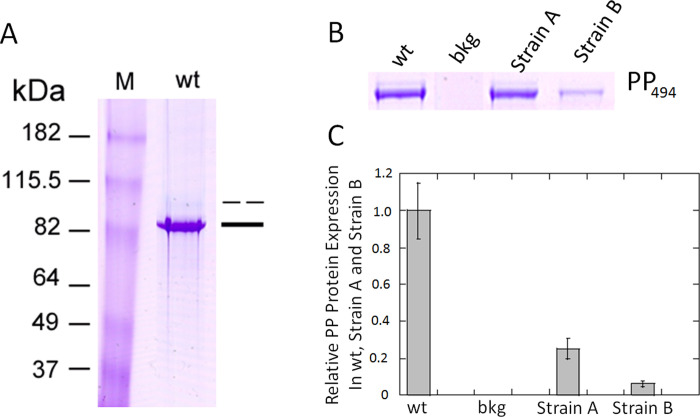
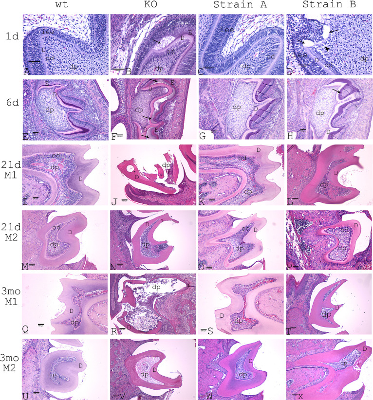
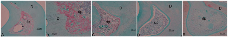
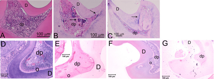
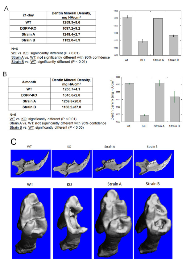
Dentin Sialoprotein (DSP) and phosphophoryn (PP) are two most dominant non-collagenous proteins in dentin, which are the cleavage products of the DSPP (dentin sialophosphoprotein) precursor protein. The absence of the DSPP gene in DSPP knock-out (KO) mice results in characteristics that are consistent with dentinogenesis imperfecta type III in humans. Symptoms include thin dentin, bigger pulp chamber with frequent pulp exposure as well as abnormal epithelial-mesenchymal interactions, and the appearance of chondrocyte-like cells in dental pulp. To better understand how DSPP influences tooth development and dentin formation, we used a bacterial artificial chromosome transgene construct (BAC-DSPP) that contained the complete DSPP gene and promoter to generate BAC-DSPP transgenic mice directly in a mouse DSPP KO background. Two BAC-DSPP transgenic mouse strains were generated and characterized. DSPP mRNA expression in BAC-DSPP Strain A incisors was similar to that from wild-type (wt) mice. DSPP mRNA expression in BAC-DSPP Strain B animals was only 10% that of wt mice. PP protein content in Strain A incisors was 25% of that found in wt mice, which was sufficient to completely rescue the DSPP KO defect in mineral density, since microCT dentin mineral density analysis in 21-day postnatal animal molars showed essentially identical mineral density in both strain A and wt mice. Strain B mouse incisors, with 5% PP expression, only partially rescued the DSPP KO defect in mineral density, as microCT scans of 21-day postnatal animal molars indicated a reduced dentin mineral density compared to wt mice, though the mineral density was still increased over that of DSPP KO. Furthermore, our findings showed that DSPP dosage in Strain A was sufficient to rescue the DSPP KO defect in terms of epithelial-mesenchymal interactions, odontoblast lineage maintenance, along with normal dentin thickness and normal mineral density while DSPP gene dosage in Strain B only partially rescued the aforementioned DSPP KO defect.


Conflict of interest statement

The authors have declared that no competing interests exist.

Figures

Fig 1
Fig 1. Diagram of mouse bacterial artificial chromosome (BAC) RP23-131C10.
This 223,760 bp BAC includes the DSP-PP gene (9.4 kb) with 149.8 kb of upstream and 64.5 kb of downstream sequences. The BAC includes DSPP gene with a complete DSPP promoter.
Fig 2
Fig 2. Examination of DSPP mRNA expression in wt, DSPP KO, BAC-DSPP transgene Strain A and Strain B teeth.
(A) A Thermo Scientific Verso cDNA kit was used to generate cDNA pools from wt, DSPP ko, Strains A and B incisor total RNAs. These cDNA pools were used to perform 30 cycles of polymerase Chain Reactions. Using a pair of PCR primers for DSPP cDNA, a 441 bp DSPP DNA band was detected in wt teeth but was not detected in DSPP KO teeth. A 233 bp glyceraldehyde-3-phosphate dehydrogenase (GAPDH) band was detected in both wt and DSPP KO teeth. (B) Using NIH image J, we determined the relative expression of normalized DSPP mRNA and present a graph of the relative expression of DSPP mRNA.
Fig 3
Fig 3. Isolation of DSP and PP from incisor extraction of wt, BAC-DSPP transgene Strain A and Strain B teeth.
(A) Lane 1, wt incisor extraction stained with Stains-All showed two bands. The lower major band stained deep blue around 82 kDa was labeled as PP (single bad). The upper minor band stained light blue was labeled as DSP (broken bar). Antibodies against DSP and PP were used to verify the identity of the incisor extract (see S1, S2 and S3 Figs). (B) Tissues were prepared as described in Methods. The incisor extracts from wt (1:10 dilution), Strain A (1:2.5 dilution), and Strain B (1:2.5 dilution) were run on a PAGE gel and stained with Stains-All. This composite image is derived from S4 Fig. (C) NIH J image bar graph of PP494 protein expression (corrected for dilutions) from Fig 3B. Since Strain A PP band derived from a 2.5x dilution of the incisor extract, showed a similar intensity of PP from 10x dilution of wt incisor extract. Thus, we concluded that wt TCA extract contain four times more PP than that of Strain A. The NIH J image then showed that Strain B contains 1/5 amount of PP compared to that of Strain A with both bands at 2.5x dilution. In summary, Strain A contained 25% of the wt PP494 protein and Stain B contained 5% of the wt PP494 protein.
Fig 4
Fig 4. H&E staining of mouse molars at various postnatal time points.
A-D: postnatal 1-day molar 1 (M1) at 400x magnification, bar for 50 μm. A: wt. B: DSPP KO. C: Strain A. D: Strain B. Arrows represent the appearance of abnormal dental pulp cells. E-H: postnatal 6-day M1. E: wt. F: DSPP KO. G: Strain A. H: Strain B. dp represents dental pulp. od represents odontoblast layer. D represents dentin. E represents Enamel. iee represents inner enamel epithelium. I-L: postnatal 21-day M1. I: wt. J: DSPP KO mouse. K: Strain A. L: Strain B. M-P: postnatal 21-day M2. M: wt. N: DSPP KO. O: Strain A. P: Strain B. Q-T: postnatal 3-month M1. Q: wt. R: DSPP KO. S: Strain A. T: Strain B. U-X represent postnatal 3-month M2. U: wt. V: DSPP KO. W: Strain A. X: Strain B. E-X: 100x magnification, bar for 100μm.dp represents dental pulp. D represents dentin.
Fig 5
Fig 5. Safranin O and fast green staining to examine acidic proteoglycan expression in DSPP KO, Strain A, Strain B and wt mice at 21-day and 3-month.
(A) 21-day DSPP KO M1 showed positive Safranin O staining. (B) 21-day Strain B M1 showed positive Safranin O staining. (C) 3-month Strain B M1 showed positive Safranin O staining. (D) 3-month Strain A showed M1 no Safranin O staining. (E) 3-month wt M1 showed no Safranin O staining. All images at 400x magnification.
Fig 6
Fig 6. Collagen type II (Col II) expression in Strain B molars.
(A) H&E staining of 21-day Strain B M1. 200x magnification. (B) Col II expression in 21-day Strain B M1. 200x magnification. (C) Col II expression in 3-month Strain B M1. 100x magnification. (D) H&E staining of 21-day Strain A M2. 100x magnification. (E) Col II expression of 21-day Strain A M2. 100x magnification. (F) H&E staining of 21-day wt M2. 100x magnification. (G) Col II expression of 21-day wt M2. 100x magnification. dp: dental pulp. D: dentin. Arrows represent Col II expression.
Fig 7
Fig 7. Analysis of mineral density and tooth structure in WT, DSPP KO, BAC-DSPP transgene Strains A and B.
(A) Mineral density of M1 dentin at 21-day. (B) Mineral density of M1 dentin at 3-month. (C) 3D reconstruction of 2D microCT images of 21-day old mice. Top panel: incisor and molars. Bottom panel: molar 1. Erosion indicated by arrows.

Similar articles

Cited by

References

    1. Linde A. Dentin Matrix Proteins: Composition and Possible Functions in Calcification. Anatomical Record. 1989;224:154–6. 10.1002/ar.1092240206 - DOI - PubMed
    1. Ritchie HH, Yee CT, Tang XN, Dong ZH, Fuller RS. DSP-PP Precursor Protein Cleavage by Tolloid-Related-1 Protein and by Bone Morphogenetic Protein-1. PLoS ONE. 2012;7(7):e41110. 10.1371/journal.pone.0041110 - DOI - PMC - PubMed
    1. Xiao S, Yu C, Chou X, Yuan W, Wang Y, Bu L, et al.. Dentinogenesis imperfecta 1 with or without progressive hearing loss associated with distinct mutations in DSPP. Nat Genet. 2001;27:201–4. 10.1038/84848 - DOI - PubMed
    1. Zhang X, Zhao J, Li C, Gao S, Qiu C, Liu P, et al.. DSPP mutation in dentinogenesis imperfecta shields type II. Nat Genet. 2001;27:151–2. 10.1038/84765 - DOI - PubMed
    1. Sreenath TL, Thyagarajan T, Hall B, Longenecker G, D’Souza RN, Hong S, et al.. Dentin sialophosphoprotein knockout mouse teeth display widened predentin zone and develop defective dentin mineralization similar to human dentiogenesis imperfecta type III. Journal of Biological Chemistry. 2003;278:24874–80. - PubMed

Publication types

MeSH terms