Positron emission tomography and magnetic resonance imaging in experimental human malaria to identify organ-specific changes in morphology and glucose metabolism: A prospective cohort study
- PMID: 34038421
- PMCID: PMC8154100
- DOI: 10.1371/journal.pmed.1003567
Positron emission tomography and magnetic resonance imaging in experimental human malaria to identify organ-specific changes in morphology and glucose metabolism: A prospective cohort study
Abstract
Background: Plasmodium vivax has been proposed to infect and replicate in the human spleen and bone marrow. Compared to Plasmodium falciparum, which is known to undergo microvascular tissue sequestration, little is known about the behavior of P. vivax outside of the circulating compartment. This may be due in part to difficulties in studying parasite location and activity in life.
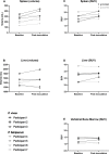
Methods and findings: To identify organ-specific changes during the early stages of P. vivax infection, we performed 18-F fluorodeoxyglucose (FDG) positron emission tomography/magnetic resonance imaging (PET/MRI) at baseline and just prior to onset of clinical illness in P. vivax experimentally induced blood-stage malaria (IBSM) and compared findings to P. falciparum IBSM. Seven healthy, malaria-naive participants were enrolled from 3 IBSM trials: NCT02867059, ACTRN12616000174482, and ACTRN12619001085167. Imaging took place between 2016 and 2019 at the Herston Imaging Research Facility, Australia. Postinoculation imaging was performed after a median of 9 days in both species (n = 3 P. vivax; n = 4 P. falciparum). All participants were aged between 19 and 23 years, and 6/7 were male. Splenic volume (P. vivax: +28.8% [confidence interval (CI) +10.3% to +57.3%], P. falciparum: +22.9 [CI -15.3% to +61.1%]) and radiotracer uptake (P. vivax: +15.5% [CI -0.7% to +31.7%], P. falciparum: +5.5% [CI +1.4% to +9.6%]) increased following infection with each species, but more so in P. vivax infection (volume: p = 0.72, radiotracer uptake: p = 0.036). There was no change in FDG uptake in the bone marrow (P. vivax: +4.6% [CI -15.9% to +25.0%], P. falciparum: +3.2% [CI -3.2% to +9.6%]) or liver (P. vivax: +6.2% [CI -8.7% to +21.1%], P. falciparum: -1.4% [CI -4.6% to +1.8%]) following infection with either species. In participants with P. vivax, hemoglobin, hematocrit, and platelet count decreased from baseline at the time of postinoculation imaging. Decrements in hemoglobin and hematocrit were significantly greater in participants with P. vivax infection compared to P. falciparum. The main limitations of this study are the small sample size and the inability of this tracer to differentiate between host and parasite metabolic activity.
Conclusions: PET/MRI indicated greater splenic tropism and metabolic activity in early P. vivax infection compared to P. falciparum, supporting the hypothesis of splenic accumulation of P. vivax very early in infection. The absence of uptake in the bone marrow and liver suggests that, at least in early infection, these tissues do not harbor a large parasite biomass or do not provoke a prominent metabolic response. PET/MRI is a safe and noninvasive method to evaluate infection-associated organ changes in morphology and glucose metabolism.
Conflict of interest statement
The authors have declared that no competing interests exist.
Figures


Similar articles
-
Evaluation of splenic accumulation and colocalization of immature reticulocytes and Plasmodium vivax in asymptomatic malaria: A prospective human splenectomy study.PLoS Med. 2021 May 26;18(5):e1003632. doi: 10.1371/journal.pmed.1003632. eCollection 2021 May. PLoS Med. 2021. PMID: 34038413 Free PMC article.
-
Positron emission tomography and magnetic resonance imaging of the brain in experimental human malaria, a prospective cohort study.Sci Rep. 2022 Apr 5;12(1):5696. doi: 10.1038/s41598-022-09748-y. Sci Rep. 2022. PMID: 35383257 Free PMC article.
-
Bone Marrow Is a Major Parasite Reservoir in Plasmodium vivax Infection.mBio. 2018 May 8;9(3):e00625-18. doi: 10.1128/mBio.00625-18. mBio. 2018. PMID: 29739900 Free PMC article.
-
Epidemiology, drug resistance, and pathophysiology of Plasmodium vivax malaria.J Vector Borne Dis. 2018 Jan-Mar;55(1):1-8. doi: 10.4103/0972-9062.234620. J Vector Borne Dis. 2018. PMID: 29916441 Free PMC article. Review.
-
Plasmodium vivax Controlled Human Malaria Infection - Progress and Prospects.Trends Parasitol. 2017 Feb;33(2):141-150. doi: 10.1016/j.pt.2016.11.001. Epub 2016 Dec 10. Trends Parasitol. 2017. PMID: 27956060 Free PMC article. Review.
Cited by
-
Frontiers in Mass Spectrometry-Based Spatial Metabolomics: Current Applications and Challenges in the Context of Biomedical Research.Trends Analyt Chem. 2024 Jun;175:117713. doi: 10.1016/j.trac.2024.117713. Epub 2024 Apr 18. Trends Analyt Chem. 2024. PMID: 40094101 Free PMC article.
-
JAK/STAT inhibition protects glucocorticoid receptor knockout mice from lethal malaria-induced hypoglycemia and hyperinflammation.EMBO Mol Med. 2025 Aug;17(8):2040-2070. doi: 10.1038/s44321-025-00264-w. Epub 2025 Jul 23. EMBO Mol Med. 2025. PMID: 40702267 Free PMC article.
-
Haematological response in experimental human Plasmodium falciparum and Plasmodium vivax malaria.Malar J. 2021 Dec 20;20(1):470. doi: 10.1186/s12936-021-04003-7. Malar J. 2021. PMID: 34930260 Free PMC article.
-
Evaluation of splenic accumulation and colocalization of immature reticulocytes and Plasmodium vivax in asymptomatic malaria: A prospective human splenectomy study.PLoS Med. 2021 May 26;18(5):e1003632. doi: 10.1371/journal.pmed.1003632. eCollection 2021 May. PLoS Med. 2021. PMID: 34038413 Free PMC article.
-
Positron emission tomography and magnetic resonance imaging of the brain in experimental human malaria, a prospective cohort study.Sci Rep. 2022 Apr 5;12(1):5696. doi: 10.1038/s41598-022-09748-y. Sci Rep. 2022. PMID: 35383257 Free PMC article.
References
-
- Marchiafava E, Bignami A. On summer-autumnal fever. London: New Sydenham. Society. 1894.
-
- Bass CC. An Attempt to Explain the Greater Pathogenicity of Plasmodium Falciparum as Compared with Other Species. Am J Trop Med Hyg. 1921;s1-1(1):29–33. 10.4269/ajtmh.1921.s1-1.29 - DOI
Publication types
MeSH terms
Substances
Associated data
LinkOut - more resources
Full Text Sources
Other Literature Sources
Medical

