

LC-MS/MS analysis of lesional and normally looking psoriatic skin reveals significant changes in protein metabolism and RNA processing
- PMID: 34038424
- PMCID: PMC8153457
- DOI: 10.1371/journal.pone.0240956
LC-MS/MS analysis of lesional and normally looking psoriatic skin reveals significant changes in protein metabolism and RNA processing
Abstract
Background: Plaque psoriasis is a chronic autoimmune disorder characterized by the development of red scaly plaques. To date psoriasis lesional skin transcriptome has been extensively studied, whereas only few proteomic studies of psoriatic skin are available.
Aim: The aim of this study was to compare protein expression patterns of lesional and normally looking skin of psoriasis patients with skin of the healthy volunteers, reveal differentially expressed proteins and identify changes in cell metabolism caused by the disease.
Methods: Skin samples of normally looking and lesional skin donated by psoriasis patients (n = 5) and samples of healthy skin donated by volunteers (n = 5) were analyzed by liquid chromatography-tandem mass spectrometry (LC-MS/MS). After protein identification and data processing, the set of differentially expressed proteins was subjected to protein ontology analysis to characterize changes in biological processes, cell components and molecular functions in the patients' skin compared to skin of the healthy volunteers. The expression of selected differentially expressed proteins was validated by ELISA and immunohistochemistry.
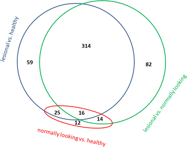
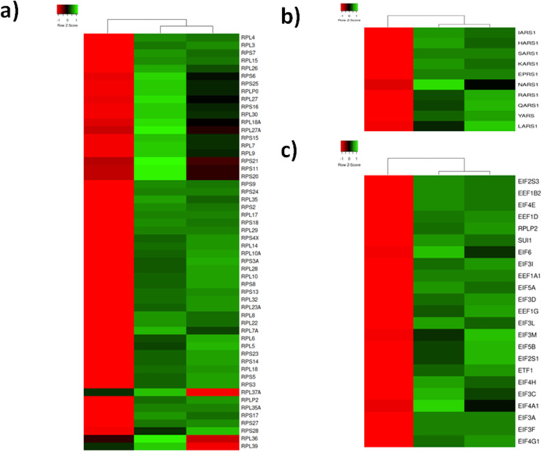
Results: The performed analysis identified 405 and 59 differentially expressed proteins in lesional and normally looking psoriatic skin compared to healthy control. In normally looking skin of the patients, we discovered decreased expression of KNG1, APOE, HRG, THBS1 and PLG. Presumably, these changes were needed to protect the epidermis from spontaneous activation of kallikrein-kinin system and delay the following development of inflammatory response. In lesional skin, we identified several large groups of proteins with coordinated expression. Mainly, these proteins were involved in different aspects of protein and RNA metabolism, namely ATP synthesis and consumption; intracellular trafficking of membrane-bound vesicles, pre-RNA processing, translation, chaperoning and degradation in proteasomes/immunoproteasomes.
Conclusion: Our findings explain the molecular basis of metabolic changes caused by disease in skin lesions, such as faster cell turnover and higher metabolic rate. They also indicate on downregulation of kallikrein-kinin system in normally looking skin of the patients that would be needed to delay exacerbation of the disease. Data are available via ProteomeXchange with identifier PXD021673.
Conflict of interest statement
The authors have declared that no competing interests exist.
Figures


Similar articles
-
Proteomic analysis of psoriatic skin tissue for identification of differentially expressed proteins: up-regulation of GSTP1, SFN and PRDX2 in psoriatic skin.Int J Mol Med. 2011 Nov;28(5):785-92. doi: 10.3892/ijmm.2011.757. Epub 2011 Jul 25. Int J Mol Med. 2011. PMID: 21805023
-
Differential Expression of Estrogen-Responsive Genes in Women with Psoriasis.J Pers Med. 2021 Sep 17;11(9):925. doi: 10.3390/jpm11090925. J Pers Med. 2021. PMID: 34575702 Free PMC article.
-
Expression Patterns of Clock Gene mRNAs and Clock Proteins in Human Psoriatic Skin Samples.Int J Mol Sci. 2021 Dec 23;23(1):121. doi: 10.3390/ijms23010121. Int J Mol Sci. 2021. PMID: 35008548 Free PMC article.
-
Analytical approaches to assess metabolic changes in psoriasis.J Pharm Biomed Anal. 2021 Oct 25;205:114359. doi: 10.1016/j.jpba.2021.114359. Epub 2021 Sep 2. J Pharm Biomed Anal. 2021. PMID: 34509137 Review.
-
Proteins in the Skin and Blood in Patients with Psoriasis: A Systematic Review of Proteomic Studies.Dermatology. 2024;240(2):317-328. doi: 10.1159/000533981. Epub 2023 Nov 7. Dermatology. 2024. PMID: 37935159
Cited by
-
Identification of gene signatures and molecular mechanisms underlying the mutual exclusion between psoriasis and leprosy.Sci Rep. 2024 Jan 25;14(1):2199. doi: 10.1038/s41598-024-52783-0. Sci Rep. 2024. PMID: 38273053 Free PMC article.
-
Analysis of PPARγ Signaling Activity in Psoriasis.Int J Mol Sci. 2021 Aug 10;22(16):8603. doi: 10.3390/ijms22168603. Int J Mol Sci. 2021. PMID: 34445309 Free PMC article.
-
Proteomic Studies of Psoriasis.Biomedicines. 2022 Mar 7;10(3):619. doi: 10.3390/biomedicines10030619. Biomedicines. 2022. PMID: 35327421 Free PMC article. Review.
-
Cross-sectional study of proteomic differences between moderate and severe psoriasis.Sci Rep. 2025 Jan 27;15(1):3387. doi: 10.1038/s41598-025-87252-9. Sci Rep. 2025. PMID: 39870771 Free PMC article.
-
Unbiased Proteomic Exploration Suggests Overexpression of Complement Cascade Proteins in Plasma from Patients with Psoriasis Compared with Healthy Individuals.Int J Mol Sci. 2024 Aug 13;25(16):8791. doi: 10.3390/ijms25168791. Int J Mol Sci. 2024. PMID: 39201477 Free PMC article.
References
MeSH terms
Substances
LinkOut - more resources
Full Text Sources
Other Literature Sources
Medical
Miscellaneous
