

Generative Adversarial Network Based Automatic Segmentation of Corneal Subbasal Nerves on In Vivo Confocal Microscopy Images
- PMID: 34038501
- PMCID: PMC8161698
- DOI: 10.1167/tvst.10.6.33
Generative Adversarial Network Based Automatic Segmentation of Corneal Subbasal Nerves on In Vivo Confocal Microscopy Images
Abstract
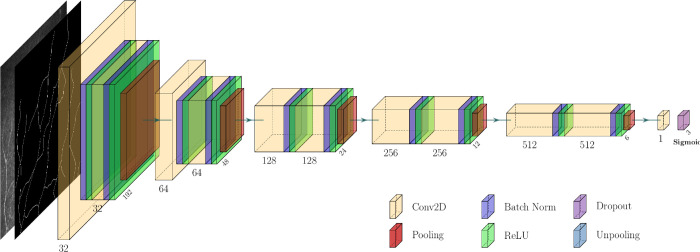
Purpose: In vivo confocal microscopy (IVCM) is a noninvasive, reproducible, and inexpensive diagnostic tool for corneal diseases. However, widespread and effortless image acquisition in IVCM creates serious image analysis workloads on ophthalmologists, and neural networks could solve this problem quickly. We have produced a novel deep learning algorithm based on generative adversarial networks (GANs), and we compare its accuracy for automatic segmentation of subbasal nerves in IVCM images with a fully convolutional neural network (U-Net) based method.
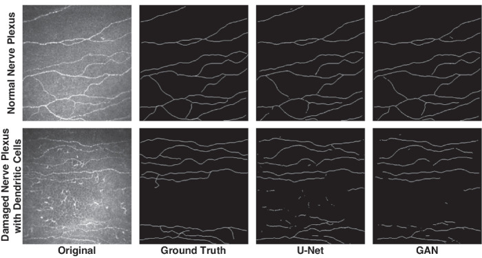
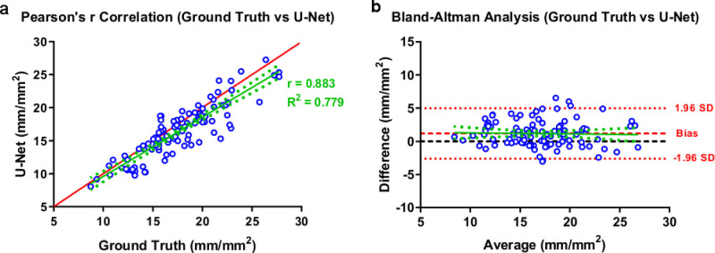
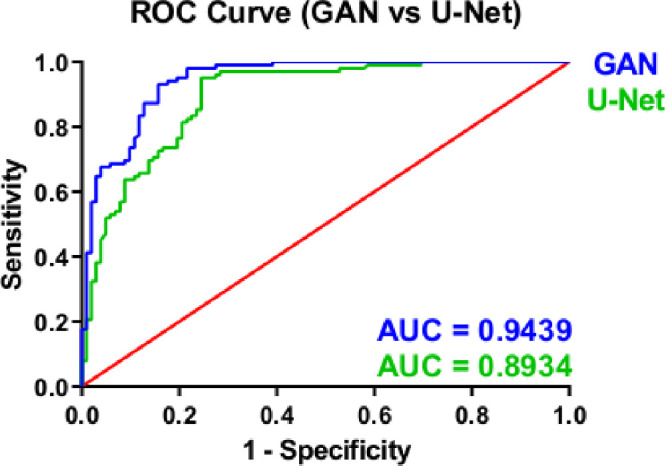
Methods: We have collected IVCM images from 85 subjects. U-Net and GAN-based image segmentation methods were trained and tested under the supervision of three clinicians for the segmentation of corneal subbasal nerves. Nerve segmentation results for GAN and U-Net-based methods were compared with the clinicians by using Pearson's R correlation, Bland-Altman analysis, and receiver operating characteristics (ROC) statistics. Additionally, different noises were applied on IVCM images to evaluate the performances of the algorithms with noises of biomedical imaging.
Results: The GAN-based algorithm demonstrated similar correlation and Bland-Altman analysis results with U-Net. The GAN-based method showed significantly higher accuracy compared to U-Net in ROC curves. Additionally, the performance of the U-Net deteriorated significantly with different noises, especially in speckle noise, compared to GAN.
Conclusions: This study is the first application of GAN-based algorithms on IVCM images. The GAN-based algorithms demonstrated higher accuracy than U-Net for automatic corneal nerve segmentation in IVCM images, in patient-acquired images and noise applied images. This GAN-based segmentation method can be used as a facilitating diagnostic tool in ophthalmology clinics.
Translational relevance: Generative adversarial networks are emerging deep learning models for medical image processing, which could be important clinical tools for rapid segmentation and analysis of corneal subbasal nerves in IVCM images.
Conflict of interest statement
Disclosure:
Figures


Similar articles
-
Segmentation and Evaluation of Corneal Nerves and Dendritic Cells From In Vivo Confocal Microscopy Images Using Deep Learning.Transl Vis Sci Technol. 2022 Jun 1;11(6):24. doi: 10.1167/tvst.11.6.24. Transl Vis Sci Technol. 2022. PMID: 35762938 Free PMC article.
-
A Deep Learning Model for Automated Sub-Basal Corneal Nerve Segmentation and Evaluation Using In Vivo Confocal Microscopy.Transl Vis Sci Technol. 2020 Jun 18;9(2):32. doi: 10.1167/tvst.9.2.32. eCollection 2020 Jun. Transl Vis Sci Technol. 2020. PMID: 32832205 Free PMC article.
-
Combining In Vivo Corneal Confocal Microscopy With Deep Learning-Based Analysis Reveals Sensory Nerve Fiber Loss in Acute Simian Immunodeficiency Virus Infection.Cornea. 2021 May 1;40(5):635-642. doi: 10.1097/ICO.0000000000002661. Cornea. 2021. PMID: 33528225 Free PMC article.
-
In Vivo Confocal Microscopy of Corneal Nerves in Health and Disease.Ocul Surf. 2017 Jan;15(1):15-47. doi: 10.1016/j.jtos.2016.09.004. Epub 2016 Oct 19. Ocul Surf. 2017. PMID: 27771327 Free PMC article. Review.
-
Combating COVID-19 Using Generative Adversarial Networks and Artificial Intelligence for Medical Images: Scoping Review.JMIR Med Inform. 2022 Jun 29;10(6):e37365. doi: 10.2196/37365. JMIR Med Inform. 2022. PMID: 35709336 Free PMC article.
Cited by
-
Potential applications of artificial intelligence in image analysis in cornea diseases: a review.Eye Vis (Lond). 2024 Mar 7;11(1):10. doi: 10.1186/s40662-024-00376-3. Eye Vis (Lond). 2024. PMID: 38448961 Free PMC article. Review.
-
Artificial-Intelligence-Enhanced Analysis of In Vivo Confocal Microscopy in Corneal Diseases: A Review.Diagnostics (Basel). 2024 Mar 26;14(7):694. doi: 10.3390/diagnostics14070694. Diagnostics (Basel). 2024. PMID: 38611606 Free PMC article. Review.
-
Segmentation and Evaluation of Corneal Nerves and Dendritic Cells From In Vivo Confocal Microscopy Images Using Deep Learning.Transl Vis Sci Technol. 2022 Jun 1;11(6):24. doi: 10.1167/tvst.11.6.24. Transl Vis Sci Technol. 2022. PMID: 35762938 Free PMC article.
-
Segmentation and Classification Approaches of Clinically Relevant Curvilinear Structures: A Review.J Med Syst. 2023 Mar 27;47(1):40. doi: 10.1007/s10916-023-01927-2. J Med Syst. 2023. PMID: 36971852 Free PMC article. Review.
-
A GAN-based deep enhancer for quality enhancement of retinal images photographed by a handheld fundus camera.Adv Ophthalmol Pract Res. 2022 Aug 19;2(3):100077. doi: 10.1016/j.aopr.2022.100077. eCollection 2022 Nov-Dec. Adv Ophthalmol Pract Res. 2022. PMID: 37846289 Free PMC article.
References
-
- Winston PH, Marvin L.. Minsky (1927–2016). Nature. 2016; 530: 282–282. - PubMed
-
- Davidovits P. Physics in biology and medicine. Waltham, MA: Academic Press; 2018.
-
- Cavanagh HD, Petroll WM, Alizadeh H, He Y-G, McCulley JP, Jester JV.. Clinical and diagnostic use of in vivo confocal microscopy in patients with corneal disease. Ophthalmology. 1993; 100: 1444–1454. - PubMed
-
- Paddock SW. Confocal microscopy: methods and protocols. New York, NY: Springer Science & Business Media; 1999.
-
- González S, Tannous Z.. Real-time, in vivo confocal reflectance microscopy of basal cell carcinoma. J Am Acad Dermatol. 2002; 47: 869–874. - PubMed
Publication types
MeSH terms
LinkOut - more resources
Full Text Sources
Other Literature Sources
