. 2021 Aug 1;162(8):bqab100.
doi: 10.1210/endocr/bqab100.

Sex-specific Disruption of the Prairie Vole Hypothalamus by Developmental Exposure to a Flame Retardant Mixture

Affiliations

Sex-specific Disruption of the Prairie Vole Hypothalamus by Developmental Exposure to a Flame Retardant Mixture

Sagi Enicole A Gillera et al. Endocrinology. .

Abstract

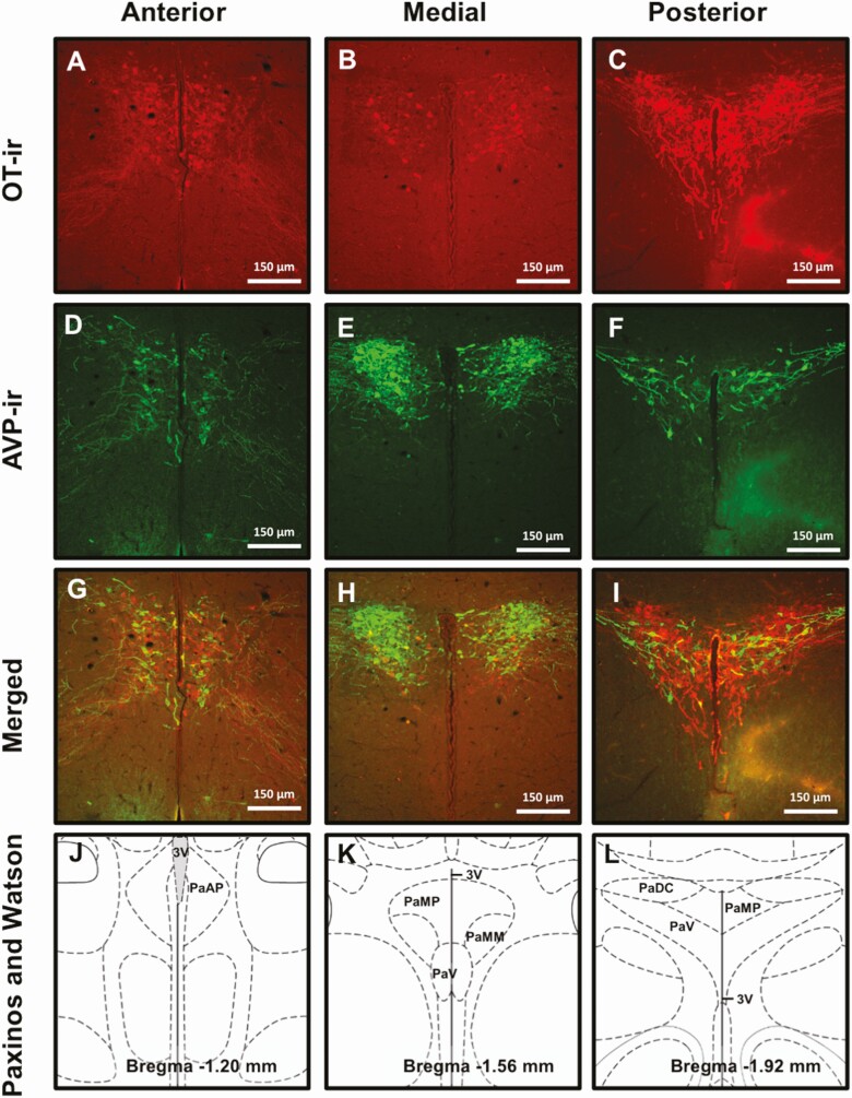
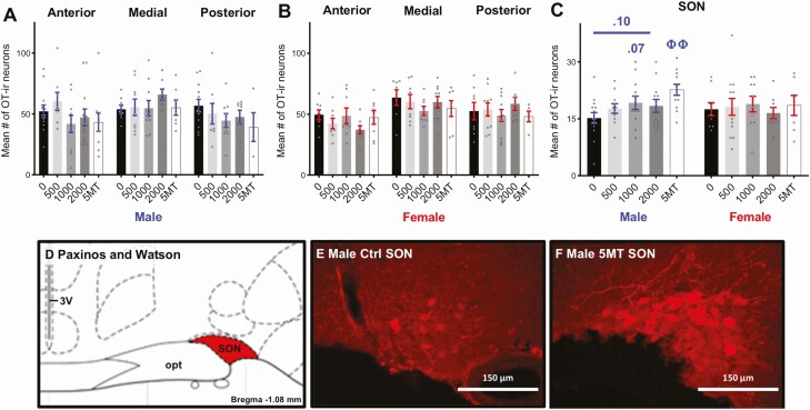
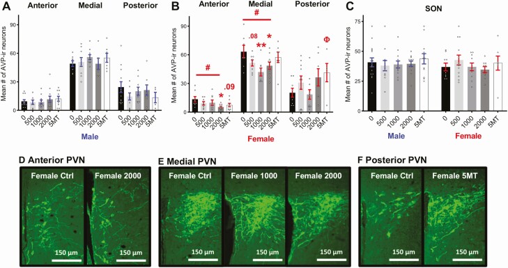
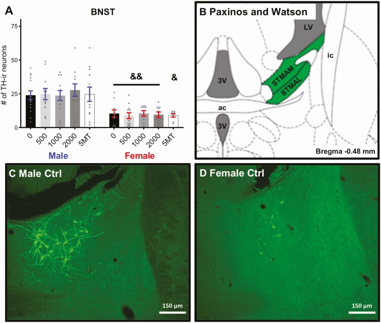
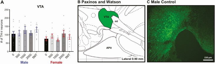
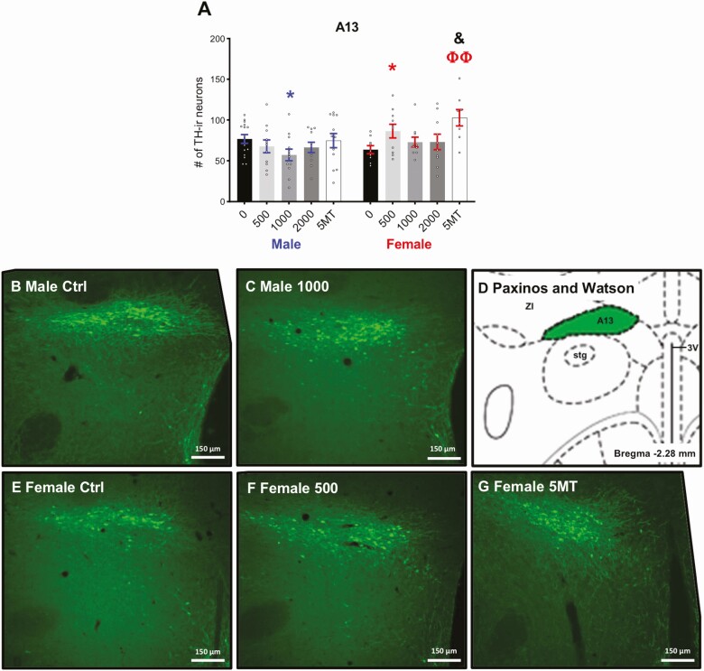
Prevalence of neurodevelopmental disorders (NDDs) with social deficits is conspicuously rising, particularly in boys. Flame retardants (FRs) have long been associated with increased risk, and prior work by us and others in multiple species has shown that developmental exposure to the common FR mixture Firemaster 550 (FM 550) sex-specifically alters socioemotional behaviors including anxiety and pair bond formation. In rats, FRs have also been shown to impair aspects of osmoregulation. Because vasopressin (AVP) plays a role in both socioemotional behavior and osmotic balance we hypothesized that AVP and its related nonapeptide oxytocin (OT) would be vulnerable to developmental FM 550 exposure. We used the prairie vole (Microtus ochrogaste) to test this because it is spontaneously prosocial. Using siblings of prairie voles used in a prior study that assessed behavioral deficits resulting from developmental FM 550 exposure across 3 doses, here we tested the hypothesis that FM 550 sex-specifically alters AVP and OT neuronal populations in critical nuclei, such as the paraventricular nucleus (PVN), that coordinate those behaviors, as well as related dopaminergic (determined by tyrosine hydroxylase (TH) immunolabeling) populations. Exposed females had fewer AVP neurons in the anterior PVN and more A13 TH neurons in the zona incerta than controls. By contrast, in FM 550 males, A13 TH neuron numbers in the zona incerta were decreased but only in 1 dose group. These results expand on previous work showing evidence of endocrine disruption of OT/AVP pathways, including to subpopulations of PVN AVP neurons that coordinate osmoregulatory functions in the periphery.

Keywords: Social; anxiety; dopamine; endocrine disrupting chemicals; endocrine disruptors; neural; neurodevelopment; oxytocin; vasopressin.


Figures

Figure 1.
Figure 1.
Representative images of each PVN region of interest (ROI) immunolabeled for OT (A-C), AVP (D-F), and both (G-I). Corresponding illustrations adapted from the Paxinos and Watson rat brain atlas depicting the landmarks and features that identify each subregion: anterior (Bregma –1.08 to –1.32 mm), medial (Bregma –1.44 to –1.80 mm), and posterior (Bregma –1.92 mm) (J-L). Scale bars, 1 μm. PaAP, paraventricular nucleus of the hypothalamus anterior parvicellular; PaDC, paraventricular nucleus of the hypothalamus dorsal cap; PaMP, paraventricular nucleus of the hypothalamus medial parvicellular; PaMM, paraventricular hypothalamic nucleus, medial magnocellular part; PaV, paraventricular nucleus ventral portion; 3V, third ventricle.
Figure 2.
Figure 2.
No significant effects of FM 550 exposure on OT-ir neurons in the PVN (A-B) or SON (C) were observed in either sex. Males exposed to 5MT had significantly more OT-ir neurons in the SON than control males (C). Illustration adapted from the Paxinos and Watson rat brain atlas depicting the landmarks used to identify the SON (Bregma –1.08 to –1.20 mm) (D). Representative images depicting increased OT-ir neuron numbers in the 5MT male SON (F) compared with control males (E). Scale bars, 150 μm. 3V, third ventricle, SON supraoptic nucleus, opt, optic tract. Bar graphs depict mean ± SEM; individual animals are depicted by circles. Significant differences due to exposure of 5MT denoted by ΦΦ (P ≤ .01).
Figure 3.
Figure 3.
No significant effects of FM 550 or 5MT exposure were observed on AVP-ir neuron numbers in male PVN (A) and SON (C). Dose dependent decreases in AVP-ir neuron numbers were observed in the anterior and medial PVN in FM 550 females (B). 5MT females had more AVP-ir neurons in the posterior PVN than control females (B). No effect of FM 550 or 5MT exposure was observed in female SON. Representative images depicting decreased AVP-ir neuron numbers in anterior (A) and medial (B) PVN of FM 550 females and increased AVP-ir neuron numbers in 5MT females (F) when compared to control females. Scale bars, 150 μm. Bar graphs depict mean ± SEM; individual animals are depicted by circles. Significant differences due to FM 550 exposure denoted by * and Φ for 5MT. #Dose–response effect within sex (*, #, and Φ P ≤ .05 and ⁎⁎P ≤ .01).
Figure 4.
Figure 4.
Males had significantly more TH-ir neurons in the BNST than females (A) and no significant effects of FM 550 or 5MT were observed in either sex. Bar graphs depict mean ± SEM; individual animals are depicted by circles. An illustration adapted from the Paxinos and Watson rat brain atlas depicting the landmarks used to identify the BNST (Bregma –0.48 mm) (B). Representative images depicting more AVP-ir neuron numbers in males (A) than females (D). Scale bars, 150 μm. Bar graphs depict mean ± SEM; individual animals are depicted by circles. Significant difference between sexes within exposure indicated by & (&&P ≤ .01). 3V, 3rd ventricle; ac, anterior commissure; STMAM, bed nucleus of the stria terminalis, medial division, anteromedial part; STMAL, bed nucleus of the stria terminalis, medial division, anterolateral part; LVe, lateral vestibular nucleus; ic, internal capsule.
Figure 5.
Figure 5.
No significant effects of FM 550 or 5MT exposure were observed on TH-ir neuron numbers in VTA (A). An illustration adapted from the Paxinos and Watson rat brain atlas depicting the landmarks used to identify the VTA (Lateral 0.90 mm) (B). Representative image of TH-ir neurons in the VTA (C). Scale bars, 150 μm. Bar graphs depict mean ± SEM; individual animals are depicted by circles.
Figure 6.
Figure 6.
Females exposed to 500 μg of FM 550 and 5MT had significantly more TH-ir A13 neurons than control females (A). There were fewer TH-ir neurons in 1000-µg males than in control males. Representative images depicting decreased TH-ir neuron numbers in the 1000-µg males (C) compared with control males (B) and increased TH-ir neuron numbers in the 500 µg FM 550 (F) and 5MT females (G) when compared with control females (E). A corresponding illustration adapted from the Paxinos and Watson rat brain atlas depicting the landmarks used to identify the BNST (Bregma –2.28 mm) (D). Bar graphs depict mean ± SEM; individual animals are depicted by circles. Significant differences due to FM 550 exposure denoted by * and Φ for 5MT. Significant difference between sexes within exposure indicated by & (* and & P ≤ .05; ΦΦ P ≤ .01). ZI, zona incerta; A13, A13 dopamine cells; Stg, sigmoid hypothalamic nucleus; 3V, 3rd ventricle.

Similar articles

Cited by

References

    1. Dufour P, Charlier C. Brominated flame retardant: environmental and exposed individuals’ health impact. Ann Biol Clin (Paris). 2017;75(2):146-157. - PubMed
    1. Xiong P, Yan X, Zhu Q, et al. A review of environmental occurrence, fate, and toxicity of novel brominated flame retardants. Environ Sci Technol. 2019;53(23):13551-13569. - PubMed
    1. Patisaul HB. Endocrine disruption of vasopressin systems and related behaviors. Front Endocrinol (Lausanne). 2017;8:134. - PMC - PubMed
    1. Landgraf R, Neumann ID. Vasopressin and oxytocin release within the brain: a dynamic concept of multiple and variable modes of neuropeptide communication. Front Neuroendocrinol. 2004;25(3-4):150-176. - PubMed
    1. Baribeau DA, Anagnostou E. Oxytocin and vasopressin: linking pituitary neuropeptides and their receptors to social neurocircuits. Front Neurosci. 2015;9:335. - PMC - PubMed

Publication types