. 2021 Nov 15;76(12):2147-2155.
doi: 10.1093/gerona/glab138.

The Indigenous South American Tsimane Exhibit Relatively Modest Decrease in Brain Volume With Age Despite High Systemic Inflammation

Affiliations

The Indigenous South American Tsimane Exhibit Relatively Modest Decrease in Brain Volume With Age Despite High Systemic Inflammation

Andrei Irimia et al. J Gerontol A Biol Sci Med Sci. .

Abstract

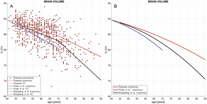
Brain atrophy is correlated with risk of cognitive impairment, functional decline, and dementia. Despite a high infectious disease burden, Tsimane forager-horticulturists of Bolivia have the lowest prevalence of coronary atherosclerosis of any studied population and present few cardiovascular disease (CVD) risk factors despite a high burden of infections and therefore inflammation. This study (a) examines the statistical association between brain volume (BV) and age for Tsimane and (b) compares this association to that of 3 industrialized populations in the United States and Europe. This cohort-based panel study enrolled 746 participants aged 40-94 (396 males), from whom computed tomography (CT) head scans were acquired. BV and intracranial volume (ICV) were calculated from automatic head CT segmentations. The linear regression coefficient estimate β^T of the Tsimane (T), describing the relationship between age (predictor) and BV (response, as a percentage of ICV), was calculated for the pooled sample (including both sexes) and for each sex. β^T was compared to the corresponding regression coefficient estimate β^R of samples from the industrialized reference (R) countries. For all comparisons, the null hypothesis β T = β R was rejected both for the combined samples of males and females, as well as separately for each sex. Our results indicate that the Tsimane exhibit a significantly slower decrease in BV with age than populations in the United States and Europe. Such reduced rates of BV decrease, together with a subsistence lifestyle and low CVD risk, may protect brain health despite considerable chronic inflammation related to infectious burden.

Keywords: Brain aging; Cardiovascular disease; Neurodegeneration.


Figures

Figure 1.
Figure 1.
(A) Cross-sectional brain volume (BV) as a percentage of intracranial volume (ICV) and its age-dependent linear trajectories in the Tsimane (red), in the reference sample of Schippling et al. (blue) and in that of Vinke et al. (black). Dots represent Tsimane participants’ BVs. Dash-dotted lines mark the range of trajectories within the confidence interval (CI) of the bootstrapping-estimated regression coefficient estimate β^ (not within the CI of the BV measurements themselves). The Tsimane and reference samples are assumed to have the same mean BV at ages 40 y, to facilitate comparison of their slopes and to obviate the divergence of trajectories as a function of age. Note the small CIs of β^ across the magnetic resonance imaging reference samples and the substantial divergence of the Tsimane CI (red) from it, particularly after age ~70 y. The null hypothesis β T = β R fails to be accepted both on the interval 40–70 y (Tsimane vs Schippling et al. (28): t3388 = −75.47, p ≃ 1.0 × 10–250, power ≃ 99%; Tsimane vs Vinke et al.: t1477 = −60.89, p ≃ 1.0 × 10–250, power ≃ 99%) and on the interval 70–95 y (Tsimane vs Vinke et al.: t1516 = −424.05, p ≃ 1.0 × 10–250, power ≃ 99%). (B) Similar to (A), comparing quadratic models of BV trajectories as a function of age in the Tsimane (red) against quadratic models of the trajectories in Schippling et al. (blue) and in Vinke et al. (29) (black). CIs and data points for individual Tsimane BV values are omitted to facilitate comparison. The curvilinear fit to the Tsimane data confirms and reproduces a slower rate of BV decrease in the Tsimane after age 70, in contrast to the reference samples, where faster rates of cross-sectional BV decline are observed.
Figure 2.
Figure 2.
Axial, sagittal, and coronal computed tomography (CT) slices for 3 representative Tsimane subjects (3 females of ages 40, 60, and 80 y, respectively). Typical aging effects upon the gray matter (GM) and white matter (WM) are highlighted, with WM depicted by semi-transparent beige overlays superimposed onto the CT scan images. GM aging features include overall shrinking of the gyri and widening of the sulci, particularly in the medial frontal lobes (axial inset arrows: cingulate sulci), lateral and medial temporal lobes (eg, Sylvian fissures and temporal opercula), parietal lobes (eg, parietal opercula), insulae (coronal inset arrows: circular sulci), and hippocampal formations (eg, choroid fissures and ambient cisterns). WM aging is highlighted by enlargement of cerebrospinal fluid structures (lateral and third ventricles) and by brain stem atrophy (sagittal inset arrows: boundary between the pons and medulla oblongata). Cerebellar atrophy (sagittal inset) reflects both GM and WM loss.
Figure 3.
Figure 3.
Neuroanatomic differences between a subject representative of the Tsimane trajectory at age 80 y (first row, cf. continuous red trace in Figure 1A) and a subject representative of the reference trajectory at the same age (second row, cf. continuous black trace in Figure 1A). Axial, sagittal, and coronal CT slices are shown for each subject. Key neuroanatomic distinctions between the 2 subjects are highlighted by arrows within each inset and include the extent of atrophy along (A) the circular sulcus of the insula, (B) the precentral and central sulci, and (C) the hippocampal formation.

References

    1. Manolio TA, Kronmal RA, Burke GL, et al. . Magnetic resonance abnormalities and cardiovascular disease in older adults. The Cardiovascular Health Study. Stroke. 1994;25:318–327. doi:10.1161/01.str.25.2.318 - DOI - PubMed
    1. Kim RE, Yun CH, Thomas RJ, et al. . Lifestyle-dependent brain change: a longitudinal cohort MRI study. Neurobiol Aging. 2018;69:48–57. doi:10.1016/j.neurobiolaging.2018.04.017 - DOI - PubMed
    1. Jefferson AL, Massaro JM, Wolf PA, et al. . Inflammatory biomarkers are associated with total brain volume: the Framingham Heart Study. Neurology. 2007;68:1032–1038. doi:10.1212/01.wnl.0000257815.20548.df - DOI - PMC - PubMed
    1. Marsland AL, Gianaros PJ, Kuan DC, Sheu LK, Krajina K, Manuck SB. Brain morphology links systemic inflammation to cognitive function in midlife adults. Brain Behav Immun. 2015;48:195–204. doi:10.1016/j.bbi.2015.03.015 - DOI - PMC - PubMed
    1. Sala-Llonch R, Idland AV, Borza T, et al. . Inflammation, amyloid, and atrophy in the aging brain: relationships with longitudinal changes in cognition. J Alzheimers Dis. 2017;58:829–840. doi:10.3233/JAD-161146 - DOI - PubMed

Publication types