. 2021 Aug;41(8):2293-2314.
doi: 10.1161/ATVBAHA.121.315957. Epub 2021 May 27.

Nrg1β Released in Remote Ischemic Preconditioning Improves Myocardial Perfusion and Decreases Ischemia/Reperfusion Injury via ErbB2-Mediated Rescue of Endothelial Nitric Oxide Synthase and Abrogation of Trx2 Autophagy

Affiliations

Nrg1β Released in Remote Ischemic Preconditioning Improves Myocardial Perfusion and Decreases Ischemia/Reperfusion Injury via ErbB2-Mediated Rescue of Endothelial Nitric Oxide Synthase and Abrogation of Trx2 Autophagy

Venkatesh Kundumani-Sridharan et al. Arterioscler Thromb Vasc Biol. 2021 Aug.

Abstract

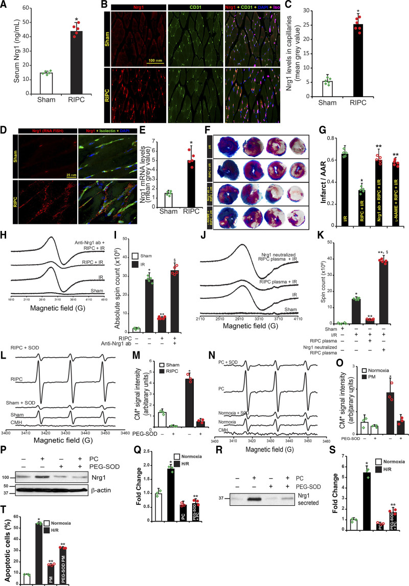
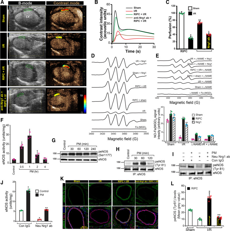
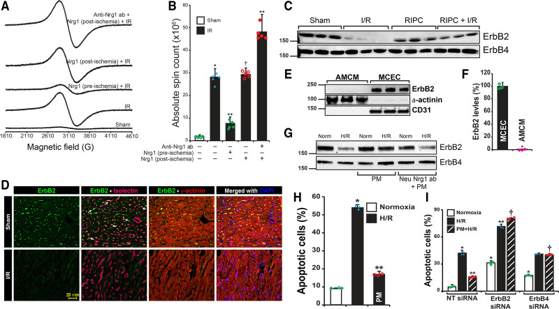
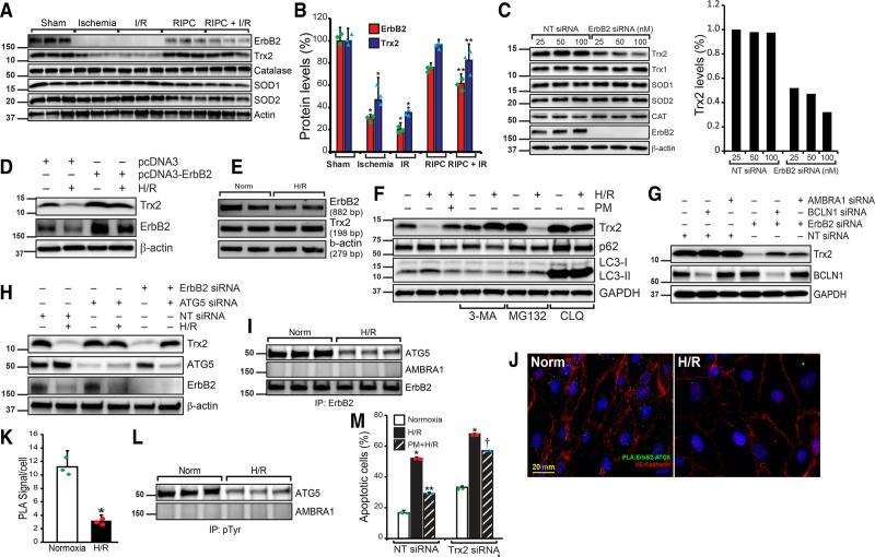
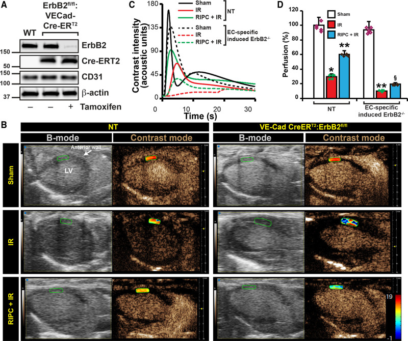
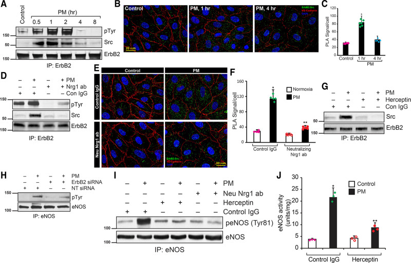
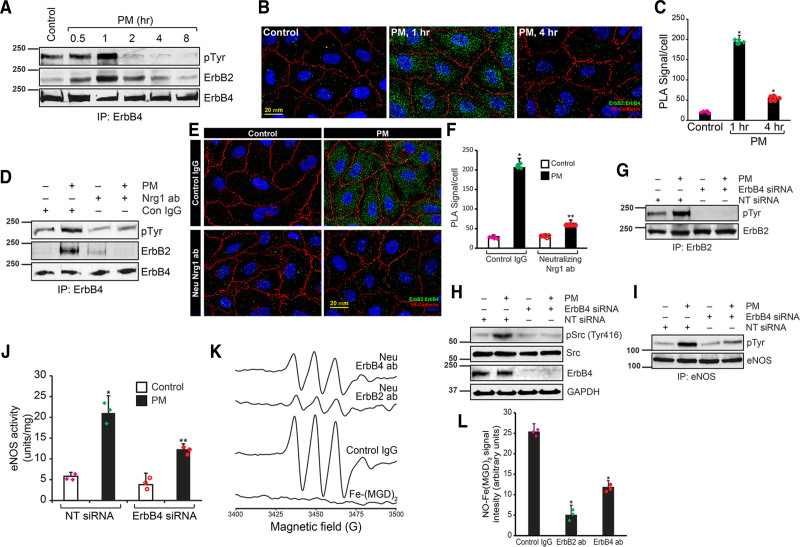
OBJECTIVE: Remote ischemic preconditioning (RIPC) is an intervention process where the application of multiple cycles of short ischemia/reperfusion (I/R) in a remote vascular bed provides protection against I/R injury. However, the identity of the specific RIPC factor and the mechanism by which RIPC alleviates I/R injury remains unclear. Here, we have investigated the identity and the mechanism by which the RIPC factor provides protection. APPROACH AND RESULTS: Using fluorescent in situ hybridization and immunofluorescence, we found that RIPC induces Nrg1β expression in the endothelial cells, which is secreted into the serum. Whereas, RIPC protected against myocardial apoptosis and infarction, treatment with neutralizing-Nrg1 antibodies abolished the protective effect of RIPC. Further, increased superoxide anion generated in RIPC is required for Nrg1 expression. Improved myocardial perfusion and nitric oxide production were achieved by RIPC as determined by contrast echocardiography and electron spin resonance. However, treatment with neutralizing-Nrg1β antibody abrogated these effects, suggesting Nrg1β is a RIPC factor. ErbB2 (Erb-B2 receptor tyrosine kinase 2) is not expressed in the adult murine cardiomyocytes, but expressed in the endothelial cells of heart which is degraded in I/R. RIPC-induced Nrg1β interacts with endothelial ErbB2 and thereby prevents its degradation. Mitochondrial Trx2 (thioredoxin) is degraded in I/R, but rescue of ErbB2 by Nrg1β prevents Trx-2 degradation that decreased myocardial apoptosis in I/R. CONCLUSIONS: Nrg1β is a RIPC factor that interacts with endothelial ErbB2 and prevents its degradation, which in turn prevents Trx2 degradation due to phosphorylation and inactivation of ATG5 (autophagy-related 5) by ErbB2. Nrg1β also restored loss of eNOS (endothelial nitric oxide synthase) function in I/R via its interaction with Src.


Figures

Figure 1.
Figure 1.
Superoxide-induced Nrg1 (neuregulin 1) release by the microvascular endothelial bed of the hindlimb acts as remote ischemic preconditioning (RIPC)-factor and protects the myocardium from ischemia/reperfusion (I/R)-induced injury. A, Mice were either subjected to sham or three cycles of 5 min hindlimb ischemia followed by 5 min reperfusion. Thirty minutes after the last I/R cycle, serum Nrg1 levels were determined by ELISA. Mean±SEM was plotted as a bar graph (n=5). *P<0.01 vs sham. B, Formaldehyde fixed paraffine embedded gastrocnemius muscle sections from sham or RIPC-treated mice were immunostained with anti-Nrg1 and anti-CD31 antibodies along with isolectin GS-IB4, scale bar=100 mm. C, Nrg1 specific fluorescent signals were quantitated, and mean±SEM fluorescent values were plotted as a bar graph (n=6). *P<0.01 vs sham (Student t test). D, To confirm the source of Nrg1, gastrocnemius muscle sections were hybridized with mouse Nrg1 Stellaris fluorescent in situ hybridization (FISH) Probe set labeled with CAL Fluor Red 590 Dye. Capillaries in the sections were stained with isolectin GS-IB4 conjugated with Alexa Fluor 488. Scale bar=25 mm. E, Mean fluorescence values from RNA FISH was quantitated, and mean±SD plotted as a bar graph (n=5). *P<0.01 vs sham. F, Mice were treated with control IgG or with neutralizing Nrg1 antibodies or l-NAME subjected to either I/R surgery or RIPC followed by I/R surgery, and TTC staining was performed. G, Infarct area in relation to area at risk (AAR) was calculated, and mean±SD plotted as a bar graph (n=5). *P<0.01 vs I/R, **P<0.01 vs RIPC+I/R (ANOVA). H, Mice were subjected to sham or myocardial I/R or RIPC followed by I/R surgery and perfused with annexin-V-Fe complex as described in Methods. The infarcted tissue was excised, and tissue bound annexin-V was quantified by measuring annexin-V conjugated paramagnetic iron by electron spin resonance (EPR). To block the function of circulating Nrg1 in mice, neutralizing anti-Nrg1 antibodies (150 µg/kg) or control IgG was administered to mice before RIPC and then subjected to RIPC+IR injury. I, Absolute spin count was calculated from EPR spectra, and means±SD (n=5) was plotted as a bar graph. *P<0.01 vs sham, **P<0.01 vs IR, §P<0.01 vs RIPC+IR, (ANOVA). J, Plasma from RIPC-subjected mice were prepared. Sibling mice were administered with RIPC, or Nrg-1 neutralized RIPC-plasma and then subjected to IR. Tissue apoptosis was measured as in H. K, Tissue bound annexin-V was quantified, and mean±SD was plotted as a bar graph (n=5). *P<0.01 vs sham, **P<0.01 vs IR, §P<0.01 vs RIPC+IR (ANOVA). L, Superoxide formation was quantitated in gastrocnemius tissue from sham or RIPC-exposed mice. Superoxide formation isolated was determined by EPR using 1-hydroxy-3-methoxycarbonyl-2,2,5,5-tetramethylpyrrolidine hydrochloride (CMH). M, CM* EPR signals were quantitated by Spin Count using Xenon nano 1.2 software and plotted as a bar graph (n=3). *P<0.01 vs sham (ANOVA). N, HMVECs were exposed to hypoxia for 2 h, followed by reoxygenation for 1 h (PC, preconditioning), and superoxide formation in HCAECs was determined by EPR using CMH. O, CM* EPR signals were quantitated and plotted as a bar graph (n=3). *P<0.01 vs normoxia (ANOVA). L–O, PEG-SOD (polyethylene glycol-superoxide dismutase) was added to determine the superoxide-specific EPR signal. P, HMVECs (n=3) were incubated with or without PEG-SOD for 6 h and subjected to PC. Cell lysates were analyzed for full-length Nrg1; (Q) Densitometry of Figure 1P, *P<0.05 vs untreated, **P<0.01 vs PC. R, Detection of Nrg1in culture medium from control or PC-subjected cells. Medium was analyzed for the full length and secreted Nrg1, respectively, by Western blotting, *P<0.01 vs HR, **P<0.01 vs PEG-SOD. T, HCAECs were incubated with preconditioned medium (PM) obtained from control or PEG-SOD treated HMVECs. Then they were exposed to hypoxia/reoxygenation (H/R). Cells undergoing apoptosis were labeled with annexin V-FITC conjugate, and the percentage of apoptotic cells was quantitated by fluorescence-activated cell sorting analysis using Attune NxT Flow Cytometer and plotted as a bar graph (n=3). *P<0.01 vs normoxia, **P<0.01 vs PM+H/R (ANOVA, Tukey post test).
Figure 2.
Figure 2.
Remote ischemic preconditioning (RIPC) improved cardiac perfusion via Nrg1 (neuregulin 1)-dependent eNOS (endothelial nitric oxide synthase) activation. A, Mice were subjected to sham or myocardial ischemia/reperfusion (I/R) or RIPC+I/R surgery, and at the end of the reperfusion period, the nontargeted contrast agent was injected to mice via femoral vein while collecting B-mode/contrast mode images of the heart from long-axis view using Vevo 3100. To block the function of circulating Nrg1 in mice, neutralizing anti-Nrg1 antibodies (150 µg/kg) was administered to mice before RIPC and then subjected to RIPC+I/R injury. B, Contrast intensities in the left ventricle (LV) anterior wall downstream to the ligation site were quantitated and plotted over time. Loss of peak height and the rightward shift of peak indicates hypoperfusion. C, From the rate of contrast agent intensity change, the perfusion index was calculated. Percentage of perfusion was calculated based on the perfusion index assuming its level in the sham group is 100% and blotted as a bar graph (n=5) and shown as a heat map over the anterior LV in A. Values are means±SEM (n=5 mice). *P<0.01 vs sham, **P<0.01 vs I/R, §P<0.01 vs RIPC+I/R (ANOVA). D, Left coronary artery from mice were isolated, and after indicated treatments, NO formation was detected by electron spin resonance (EPR) spectrometry using NO spin trap Fe2+-(N-methyl-D-glucamine dithiocarbamate) 2 (Fe-[MGD]2) as described in methods. E, NO-Fe(MGD) 2 spin count was quantified and plotted as a bar graph (n=5). *P<0.01 vs sham; **P<0.01 vs I/R (ANOVA). F–H, HMVECs were exposed to 2 h hypoxia followed by 1-hour reoxygenation, and the medium was collected (preconditioned medium [PM]). HCAECs were treated with PM for the indicated period, lysed, and cell lysates were collected. An equal amount of cell lysates were analyzed for eNOS activity (F) and plotted as a bar graph (n=3). *P<0.01 vs control (ANOVA). An equal amount of cell lysates were analyzed for eNOS phosphorylation at Ser 1177 by Western blotting using its phospho-specific antibodies and normalized with total eNOS levels (G), or the cell lysates were immunoprecipitated with anti-eNOS antibodies, and the immunoprecipitates were analyzed by Western blotting using anti-phospho-tyrosine specific antibodies (H). I–J, To determine the role of HMVEC-released Nrg1 in PM in the activation of eNOS, the collected PM was pretreated with neutralizing anti-Nrg1 antibodies and incubated with human coronary artery endothelial cell (HCAEC) for 1 h, and cell lysates were collected and analyzed for eNOS tyrosine phosphorylation by Western blotting (I) or eNOS activity (J). K, Mice were injected control IgG or neutralizing anti-Nrg1 antibodies via the tail vein. After 24 h, they were subjected to hindlimb RIPC followed by myocardial I/R, and heart from these mice was sectioned below the LAD ligation point and analyzed for eNOS phosphorylation at Tyr81 by immunofluorescence staining. Scale bar=50 mm. L, Green fluorescence localized to the coronary artery endothelial cells were quantitated (n=5), and the results were plotted as a bar graph. *P<0.05 vs I/R, **P<0.05 vs control IgG+I/R (ANOVA).
Figure 3.
Figure 3.
Only preischemic and not postischemic Nrg1β (neuregulin 1) administration protects myocardium due to loss of endothelial-ErbB2 (Erb-B2 receptor tyrosine kinase 2) during ischemia. A, To determine the protective function of Nrg1 during ischemia or reperfusion, recombinant Nrg1β (4 µg/kg) was injected into mice before ischemia (preischemia) or after ischemia (postischemia) but before reperfusion. After ischemia/reperfusion (I/R), the heart was isolated and perfused with annexin-V-Fe complex. The infarcted tissue was excised, and tissue bound annexin-V was quantified by electron spin resonance (EPR). B, The absolute spin count was calculated from the EPR spectra of paramagnetic-Fe bound to annexin-V and plotted as a bar graph. Values are means±SD (n=5 mice). *P<0.01 vs sham; **P<0.01 vs I/R, †P<0.01 vs Nrg1 (preischemic) plus IR (ANOVA). C, Mice were subjected to sham, I/R, remote ischemic preconditioning (RIPC) or RIPC+I/R, and the infarcted tissue region was excised. Protein extract was prepared from the excised tissue and analyzed by Western blotting for ErbB2 and ErbB4 levels. D, Sections of sham and I/R mouse heart were subjected to immunofluorescence staining using anti-ErbB2 and anti-α-actinin (cardiomyocyte marker) antibodies. Isolectin-Alexa Fluor 647 conjugate was used to stain endothelial cells selectively. Fluorescent images of the stained sections were obtained using an upright Zeiss fluorescence microscope (AxioImager Z2) via 40×/1.4 NA objective. Scale bar=20 mm. E and F, Adult mouse cardiomyocytes and mouse cardiac endothelial cells were isolated from adult mouse heart, and cell lysate was prepared. An equal amount of protein from cell lysates was analyzed for ErbB2 levels by Western blotting. Blot was reprobed for α-actinin and CD31 (F). ErbB2 levels were quantified and plotted as a bar graph (n=3). *P<0.01 vs MCEC (Student t test). G, HCAECs were pretreated with preconditioned medium (PM) or Nrg1-neutralized PM, then exposed to hypoxia/reoxygenation (H/R), and cell lysates were prepared. An equal amount of protein from each sample was analyzed for ErbB2 and ErbB4 levels by Western blotting. H, HCAECs were pretreated with or without PM and exposed to H/R. At the end of treatment, cells undergoing apoptosis were labeled with annexin V-FITC conjugate, and the percentage of apoptotic cells was quantitated by fluorescence-activated cell sorting analysis using Attune NxT Flow Cytometer and plotted as a bar graph (n=3). *P<0.01 vs normoxia, **P<0.01 vs H/R (ANOVA). I, HCAECs were transfected with nontarget (NT) or ErbB4 or ErbB4 siRNA (100 nmol/L), and after recovery from transfection, they were treated with or without PM and exposed to H/R. Cells undergoing apoptosis were labeled with annexin V-FITC conjugate, and the percentage of apoptotic cells was quantitated and plotted as a bar graph (n=3). *P<0.01 vs NT siRNA normoxia, **P<0.01 vs NT siRNA H/R; †P<0.01 vs NT siRNA PM+H/R (ANOVA). ANOVA followed by Tukey post test was performed using GraphPad-Prism software (version 8).
Figure 4.
Figure 4.
Remote ischemic preconditioning (RIPC) protects ErbB2 (Erb-B2 receptor tyrosine kinase 2) from ischemia/reperfusion (I/R)-mediated degradation and the loss of ErbB2 results in Trx2 (thioredoxin 2) autophagy and apoptosis. A, Mice were subjected to a sham procedure, 30 min ischemia, I/R, RIPC or RIPC+I/R, and the infarcted tissue region were excised. Protein extract was prepared from the excised tissue and analyzed by Western blotting for ErbB2, Trx2, catalase, SOD1, SOD2, and total actin. B, ErbB2 and Trx2 levels in the blots were quantified and plotted as a bar graph (n=3). *P<0.01 vs sham; **P<0.01 vs I/R. C, HCAECs were transfected with nontarget (NT) or ErbB2 siRNA (25, 50, 100 nmol/L). After 48 h of transfection, cell lysates were prepared, and an equal amount of protein from each cell lysate was analyzed for Trx1, Trx2, SOD1, SOD2, and catalase levels using their specific antibodies. Blots were reprobed with anti-ErbB2 and anti-β-actin antibodies to determine the depletion of ErbB2 and equal protein loading, respectively. Bar graph shows the level of Trx2. D, HCAECs were transfected with pcDNA3 empty or pcDNA3-ErbB2 plasmids. After 24 h of transfection, cells were exposed to hypoxia/reoxygenation (H/R), and cell lysates were prepared. An equal amount of protein from each sample was analyzed for Trx2 and ErbB2 levels using their specific antibodies. Blots were reprobed with anti-β-actin antibodies. E, HCAECs were exposed to normoxia or H/R, mRNA was isolated, and cDNA was synthesized. PCR was performed to determine levels of ErbB2 (primers, 5′- GTGCTGGTCAAGAGTCCCAACCATG-3′ and 5′- ATCTGGCTGGTTCACATATTCAGGC-3’), Trx2 (primers, 5′- CCACACCTTGGTCCTCATCT-3′ and 5′- AGGAGGTGGAAGGGATGACT-3′), and β-actin (primers, 5′-CCGCCAGCTCACCAT-3′ and 5′-GTGTGGTGCCAGATTTTCTC-3′). F, HCAECs were treated with 3-methyl adenine (3-MA), MG132 (proteasomal inhibitor), or chloroquine (CLQ) before exposing them to H/R. After the treatment, cells were lysed, and the cell lysates were analyzed by Western blotting for Trx2, p62, microtubule-associated protein 1, light chain 3-I/II level using its specific antibody, and the blot was reprobed for β-actin for normalization. G, HCAECs were transfected with Ambra1 or beclin 1 (BCLN1) siRNA in combination with NT or ErbB2 siRNA (100 nmol/L). After 48 h of transfection, cell lysates were prepared, and an equal amount of protein from each cell lysate was analyzed for Trx2, beclin 1, and GAPDH levels using their specific antibodies. H, HCAECs were transfected with NT or ATG5 (autophagy-related 5) siRNA and exposed to H/R or transfected with ATG5 siRNA and ErbB2 siRNA. After 48 h of transfection and H/R treatment, cell lysates were prepared, and an equal amount of protein from each cell lysate was analyzed for Trx2, ATG5, ErbB2, and GAPDH levels using their specific antibodies. I, HCAECs were exposed to normoxia or H/R, and cell lysates were immunoprecipitated with anti-ErbB2 antibodies. Immunoprecipitates were analyzed for ATG5, AMBRA1 (activating molecule in beclin 1-regulated autophagy), and ErbB2. J, To study the in situ interaction of ErbB2 and ATG5, HCAECs were subjected to H/R and proximity ligation assay (PLA) was performed using anti-ErbB2 and anti-ATG5 antibodies. Scale bar=20 mm. K, Green foci-proximity signals of ErbB2 and ATG5 association were counted and plotted as a bar graph. *P<0.01 vs normoxia. Student t test. L, HCAECs were exposed to normoxia or H/R, and cell lysates were immunoprecipitated with anti-pTyr antibodies. Immunoprecipitates were analyzed for ATG5 and AMBRA1. M, HCAECs were transfected with NT or Trx2 siRNA (100 nmol/L), and after recovery from transfection, they were treated with or without preconditioned medium (PM) and exposed to H/R. Cells undergoing apoptosis were labeled with annexin V-FITC conjugate, and the percentage of apoptotic cells was quantitated and plotted as a bar graph (n=3). *P<0.01 vs NT siRNA normoxia, **P<0.01 vs NT siRNA H/R, †P<0.01 vs NT siRNA PM+H/R. One way ANOVA was performed using GraphPad-Prism software (version 8).
Figure 5.
Figure 5.
Endothelial-specific deletion of ErbB2 (Erb-B2 receptor tyrosine kinase 2) results in loss of remote ischemic preconditioning (RIPC)-mediated protection of cardiac perfusion. A, To generate a mouse model of endothelial-specific inducible deletion of ErbB2, we crossed ErbB2 floxed mice with VECad-Cre-ERT2 mice. To test the endothelial-specific deletion of ErbB2, we isolated endothelial cells from WT and ErbB2fl/fl:VECad-Cre-ERT2 mice and treated with tamoxifen (30 µg/mL) for 48 h. After the treatment, cells were lysed, and an equal amount of protein from cell lysates was analyzed for ErbB2 levels by Western blotting using anti-ErbB2 antibodies. Blots were reprobed with anti-Cre, anti-CD31, and anti-β-actin antibodies. B, To induce deletion of ErbB2 in endothelial cells, ErbB2fl/fl:VECad-Cre-ERT2 mice were injected with three doses of tamoxifen (75 mg/kg). After the induced deletion, ErbB2fl/fl:VECad-Cre-ERT2 and nontarget (NT) mice were subjected to sham or myocardial ischemia/reperfusion (I/R) or RIPC+I/R surgery. At the end of the reperfusion period, the nontargeted contrast agent was injected into mice via the left femoral vein while collecting B-mode/contrast mode images of the heart from the long-axis view using Vevo 3100. C, Contrast intensities in the left ventricle (LV) anterior wall downstream to the ligation site were quantitated and plotted over time. D, From the rate of contrast agent intensity change, percentage of perfusion was calculated based on the perfusion index assuming its level in the sham group is 100% and blotted as a bar graph (n=5) and shown as a heat map over the anterior LV in A. Values are means±SEM (NT, n=5; ErbB2fl/fl:VECad-Cre-ERT2, n=3). *P<0.01 vs NT sham, **P<0.01 vs NT I/R, §P<0.01 vs NT RIPC+I/R (ANOVA).
Figure 6.
Figure 6.
Endothelial-ErbB2 (Erb-B2 receptor tyrosine kinase 2) directly associates with Src and activate eNOS (endothelial nitric oxide synthase). A, Human coronary artery endothelial cells (HCAECs) were treated with preconditioned medium (PM) for the indicated period, and cell lysates were prepared. An equal amount of protein from cell lysates was immunoprecipitated using the anti-ErbB2 antibody. The immunoprecipitates were analyzed by Western blotting for tyrosine phosphorylation of ErbB2, level of associated Src, ErbB4, and ErbB2 using their specific antibodies. B, HCAECs were treated with CM for 1 or 4 h, and proximity ligation assay (PLA) was performed using anti-ErbB2 and anti-Src antibodies. Scale bar=20 mm. C, PLA signals were counted and plotted as a bar graph (n=5). *P<0.01 vs control (ANOVA). D, To determine the role of Nrg1 (neuregulin 1) released by HMVEC in PM, PM was pretreated with neutralizing anti-Nrg1 and then incubated with HCAECs for 1 h. At the end of treatment, cell lysates were prepared and analyzed for ErbB2 tyrosine phosphorylation and its interaction with Src immunoprecipitation followed by Western blotting. E, HCAECs were incubated with PM or Nrg1 neutralized-PM for 1 h. and PLA was performed using anti-ErbB2 and anti-Src antibodies. Scale bar=20 mm. F, PLA signals were counted and plotted as a bar graph (n=5). *P<0.01 vs control (ANOVA). G, HCAECs were pretreated with Herceptin and incubated with PM for 1 h, and cell lysates were prepared. An equal amount of proteins from cell lysates were immunoprecipitated with anti-ErbB2 antibodies, and the immunocomplexes were analyzed for associated Src by Western blotting. H, HCAEC were transfected with ErbB2 siRNA, treated with PM for 1 h, and cell lysates were prepared and analyzed for eNOS tyrosine phosphorylation. I, HCAECs were pretreated with control IgG, Herceptin, or Nrg1 neutralizing antibodies and then incubated with PM for 1 h, and cell lysates were prepared. An equal amount of proteins from cell lysates were analyzed for eNOS-Tyr81 phosphorylation by Western blotting using its specific antibodies. J, HCAECs were pretreated with control IgG or Herceptin and incubated with PM for 1 h, and cell lysates were prepared. An equal amount of protein from cell lysates was analyzed for eNOS activity and plotted as a bar graph. *P<0.01 vs control IgG, **P<0.01 vs control IgG+PM (ANOVA).
Figure 7.
Figure 7.
Endothelial ErbB2 (Erb-B2 receptor tyrosine kinase 2) dimerizes with ErbB4, recruits Src and mediates Nrg1 (neuregulin 1)-dependent eNOS (endothelial nitric oxide synthase) activation during ischemic preconditioning. A, HCAECs were treated with preconditioned medium (PM) for the indicated period, and cell lysates were prepared. An equal amount of protein from cell lysates was immunoprecipitated using anti-ErbB4 antibody, and the immunoprecipitates were analyzed by Western blotting for tyrosine phosphorylation of ErbB4 and associated ErbB2 levels using their specific antibodies. B, To study the in vivo interaction of ErbB2 and ErbB4, HCAECs were treated with PM for 1 or 4 h, and proximity ligation assay (PLA) was performed using anti-ErbB2 and anti-ErbB4 antibodies. Scale bar=20 mm. C, Green foci-proximity signals of ErbB2 and ErbB2 association were counted and plotted as a bar graph. *P<0.01 vs control (ANOVA). D–F, HCAECs were incubated with PM or Nrg1 neutralized-PM for 1 h, and either cell lysates were prepared and analyzed for ErbB4 tyrosine phosphorylation and its interaction with ErbB2 by Western blotting as described in A or subjected to PLA (E) using anti-ErbB2 and anti-ErbB4 antibodies as described in B. PLA signals were counted and plotted as a bar graph (n=5), Scale bar=20 mm (F). *P<0.01 vs control IgG; **P<0.01 vs neutralizing anti-Nrg1 antibody+PM. G–J, HCAECs were transfected with ErbB4 siRNA, treated with PM for 1 h., and the cell lysates were prepared and analyzed for ErbB2 tyrosine phosphorylation (G), Src activation (H), or eNOS tyrosine phosphorylation (I). An equal amount of protein from cell lysates was analyzed for eNOS activity (J). *P<0.01 vs nontarget (NT) siRNA; **P<0.01 vs NT siRNA+PM (ANOVA). K, To study the role of ErbB2 and ErbB4 in NO formation in coronary arteries, left coronary artery were isolated and incubated with control IgG, neutralizing anti-ErbB2 or neutralizing anti-ErbB4 antibodies, and NO release in response to ACh (10 µmol/L) was determined by electron spin resonance (EPR) using Fe-(MGD)2. L, Spin count of the NO-Fe(MGD)2 was calculated from EPR signals were plotted as a bar graph. *P<0.01 vs control IgG (ANOVA).

Comment in

References

    1. Hausenloy DJ, Yellon DM. Myocardial ischemia-reperfusion injury: a neglected therapeutic target. J Clin Invest. 2013; 123:92–100. doi: 10.1172/JCI62874 - PMC - PubMed
    1. Hausenloy DJ, Yellon DM. Ischaemic conditioning and reperfusion injury. Nat Rev Cardiol. 2016; 13:193–209. doi: 10.1038/nrcardio.2016.5 - PubMed
    1. Cung TT, Morel O, Cayla G, Rioufol G, Garcia-Dorado D, Angoulvant D, Bonnefoy-Cudraz E, Guérin P, Elbaz M, Delarche N, et al. . Cyclosporine before PCI in patients with acute myocardial infarction. N Engl J Med. 2015; 373:1021–1031. doi: 10.1056/NEJMoa1505489 - PubMed
    1. Heusch G. Molecular basis of cardioprotection: signal transduction in ischemic pre-, post-, and remote conditioning. Circ Res. 2015; 116:674–699. doi: 10.1161/CIRCRESAHA.116.305348 - PubMed
    1. Anttila V, Haapanen H, Yannopoulos F, Herajärvi J, Anttila T, Juvonen T. Review of remote ischemic preconditioning: from laboratory studies to clinical trials. Scand Cardiovasc J. 2016; 50:355–361. doi: 10.1080/14017431.2016.1233351 - PubMed

Publication types

MeSH terms