Inhibition of herpes simplex virus-1 infection by MBZM-N-IBT: in silico and in vitro studies
- PMID: 34039377
- PMCID: PMC8157732
- DOI: 10.1186/s12985-021-01581-5
Inhibition of herpes simplex virus-1 infection by MBZM-N-IBT: in silico and in vitro studies
Abstract
Introduction: The emergence of drug resistance and cross-resistance to existing drugs has warranted the development of new antivirals for Herpes simplex viruses (HSV). Hence, we have designed this study to evaluate the anti-viral activity of 1-[(2-methyl benzimidazole-1-yl) methyl]-2-oxo-indolin-3-ylidene] amino] thiourea (MBZM-N-IBT), against HSV-1.
Method: Molecular docking was performed to assess the affinity of MBZM-N-IBT for HSV-1 targets. This was validated by plaque assay, estimation of RNA and protein levels as well as time of addition experiments in vitro.
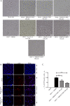
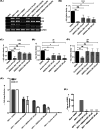
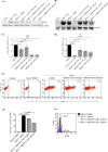
Result: Molecular docking analysis suggested the inhibitory capacity of MBZM-N-IBT against HSV-1. This was supported by the abrogation of the HSV-1 infectious viral particle formation with the IC50 value of 3.619 µM. Viral mRNA levels were also reduced by 72% and 84% for UL9 and gC respectively. MBZM-N-IBT also reduced the protein synthesis for gC and ICP8 significantly. While mRNA of ICP8 was not significantly affected, its protein synthesis was reduced by 47%. The time of addition experiment revealed the capacity of MBZM-N-IBT to inhibit HSV-1 at early as well as late stages of infection in the Vero cells. Similar effect of MBZM-N-IBT was also noticed in the Raw 264.7 and BHK 21 cells after HSV-1 infection. Supported by the in silico data, this can be attributed to possible interference with multiple HSV targets including the ICP8, ICP27, UL42, UL25, UL15 and gB proteins.
Conclusion: These results along with the lack of acute oral toxicity and significant anti-inflammatory effects suggest its suitability for further evaluation as a non-nucleoside inhibitor of HSV.
Keywords: Herpes simplex virus-1; ICP8; MBZM-N-IBT; UL9; gC.
Conflict of interest statement
The authors declare that they have no competing of interest.
Figures


Similar articles
-
MBZM-N-IBT, a Novel Small Molecule, Restricts Chikungunya Virus Infection by Targeting nsP2 Protease Activity In Vitro, In Vivo, and Ex Vivo.Antimicrob Agents Chemother. 2022 Jul 19;66(7):e0046322. doi: 10.1128/aac.00463-22. Epub 2022 Jun 29. Antimicrob Agents Chemother. 2022. PMID: 35766508 Free PMC article.
-
Inhibition of Chikungunya Virus Replication by 1-[(2-Methylbenzimidazol-1-yl) Methyl]-2-Oxo-Indolin-3-ylidene] Amino] Thiourea(MBZM-N-IBT).Sci Rep. 2016 Feb 4;6:20122. doi: 10.1038/srep20122. Sci Rep. 2016. PMID: 26843462 Free PMC article.
-
Antiviral activity of arbidol hydrochloride against herpes simplex virus I in vitro and in vivo.Int J Antimicrob Agents. 2018 Jan;51(1):98-106. doi: 10.1016/j.ijantimicag.2017.09.001. Epub 2017 Sep 7. Int J Antimicrob Agents. 2018. PMID: 28890393
-
Network Pharmacology, Molecular Docking and in vivo-based Analysis on the Effects of the MBZM-N-IBT for Arthritis.Curr Comput Aided Drug Des. 2025;21(2):194-210. doi: 10.2174/0115734099307360240731052835. Curr Comput Aided Drug Des. 2025. PMID: 39108124
-
Identification of a divalent metal cation binding site in herpes simplex virus 1 (HSV-1) ICP8 required for HSV replication.J Virol. 2012 Jun;86(12):6825-34. doi: 10.1128/JVI.00374-12. Epub 2012 Apr 4. J Virol. 2012. PMID: 22491472 Free PMC article.
Cited by
-
Antiherpetic Activity of a Root Exudate from Solanum lycopersicum.Microorganisms. 2024 Feb 11;12(2):373. doi: 10.3390/microorganisms12020373. Microorganisms. 2024. PMID: 38399777 Free PMC article.
-
MBZM-N-IBT, a Novel Small Molecule, Restricts Chikungunya Virus Infection by Targeting nsP2 Protease Activity In Vitro, In Vivo, and Ex Vivo.Antimicrob Agents Chemother. 2022 Jul 19;66(7):e0046322. doi: 10.1128/aac.00463-22. Epub 2022 Jun 29. Antimicrob Agents Chemother. 2022. PMID: 35766508 Free PMC article.
-
Conjugates of ibuprofen inhibit CHIKV infection and inflammation.Mol Divers. 2024 Jun;28(3):1261-1272. doi: 10.1007/s11030-023-10654-2. Epub 2023 Apr 21. Mol Divers. 2024. PMID: 37085737
-
HerpDock: A GUI-based gateway to HSV-1 molecular docking insights.Comput Struct Biotechnol J. 2024 Oct 13;23:3692-3701. doi: 10.1016/j.csbj.2024.10.013. eCollection 2024 Dec. Comput Struct Biotechnol J. 2024. PMID: 39507821 Free PMC article.
-
Salicylic Acid Conjugate of Telmisartan Inhibits Chikungunya Virus Infection and Inflammation.ACS Omega. 2023 Dec 21;9(1):146-156. doi: 10.1021/acsomega.3c00763. eCollection 2024 Jan 9. ACS Omega. 2023. PMID: 38222605 Free PMC article.
References
Publication types
MeSH terms
Substances
LinkOut - more resources
Full Text Sources
Other Literature Sources
Medical
Miscellaneous

