. 2021 May 26;40(1):177.
doi: 10.1186/s13046-021-01973-z.

Exosomal DLX6-AS1 from hepatocellular carcinoma cells induces M2 macrophage polarization to promote migration and invasion in hepatocellular carcinoma through microRNA-15a-5p/CXCL17 axis

Affiliations

Exosomal DLX6-AS1 from hepatocellular carcinoma cells induces M2 macrophage polarization to promote migration and invasion in hepatocellular carcinoma through microRNA-15a-5p/CXCL17 axis

Lin-Pei Wang et al. J Exp Clin Cancer Res. .

Retraction in

Abstract

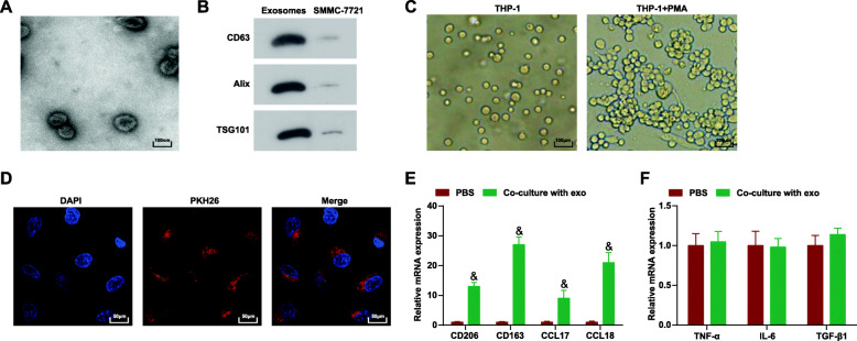
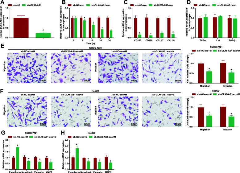
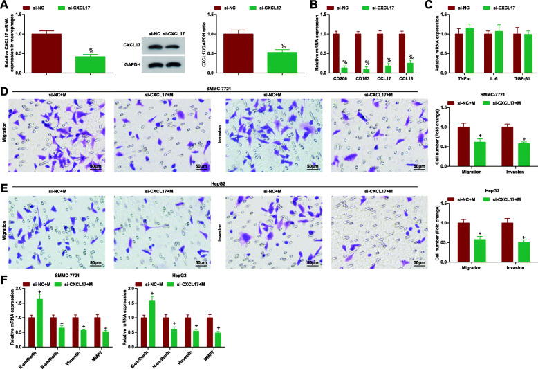
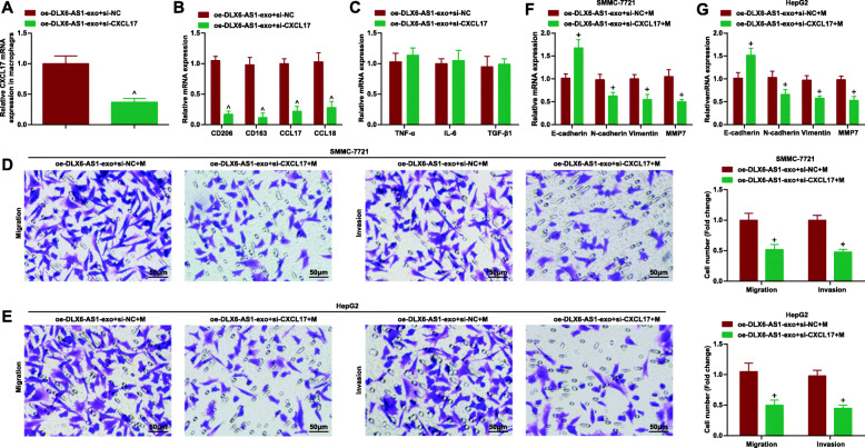
Background: Hepatocellular carcinoma (HCC) cells-secreted exosomes (exo) could stimulate M2 macrophage polarization and promote HCC progression, but the related mechanism of long non-coding RNA distal-less homeobox 6 antisense 1 (DLX6-AS1) with HCC-exo-mediated M2 macrophage polarization is largely ambiguous. Thereafter, this research was started to unearth the role of DLX6-AS1 in HCC-exo in HCC through M2 macrophage polarization and microRNA (miR)-15a-5p/C-X-C motif chemokine ligand 17 (CXCL17) axis.

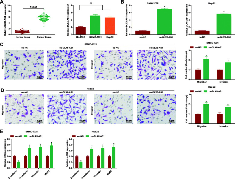
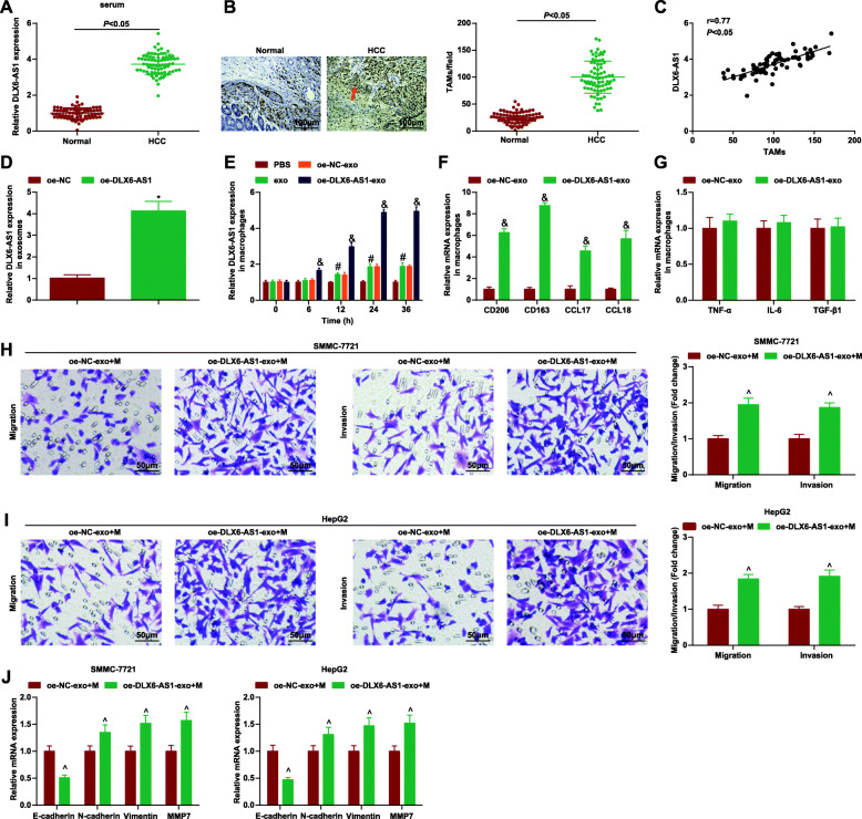
Methods: DLX6-AS1, miR-15a-5p and CXCL17 expression in HCC tissues and cells were tested. Exosomes were isolated from HCC cells with overexpressed DLX6-AS1 and co-cultured with M2 macrophages. MiR-15a-5p/CXCL17 down-regulation assays were performed in macrophages. The treated M2 macrophages were co-cultured with HCC cells, after which cell migration, invasion and epithelial mesenchymal transition were examined. The targeting relationships between DLX6-AS1 and miR-15a-5p, and between miR-15a-5p and CXCL17 were explored. In vivo experiment was conducted to detect the effect of exosomal DLX6-AS1-induced M2 macrophage polarization on HCC metastasis.

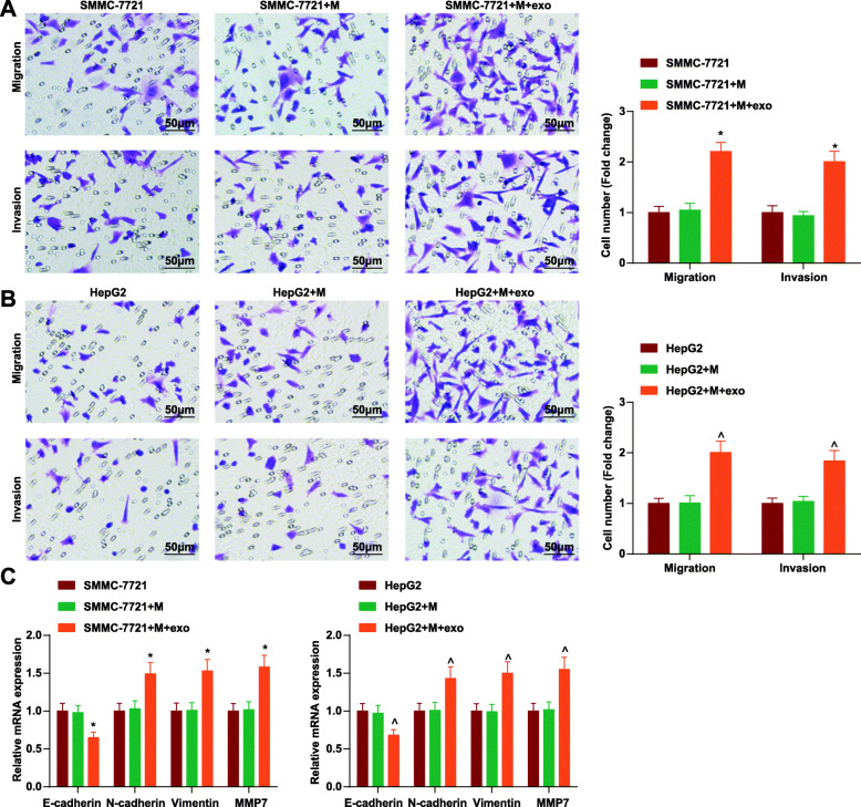
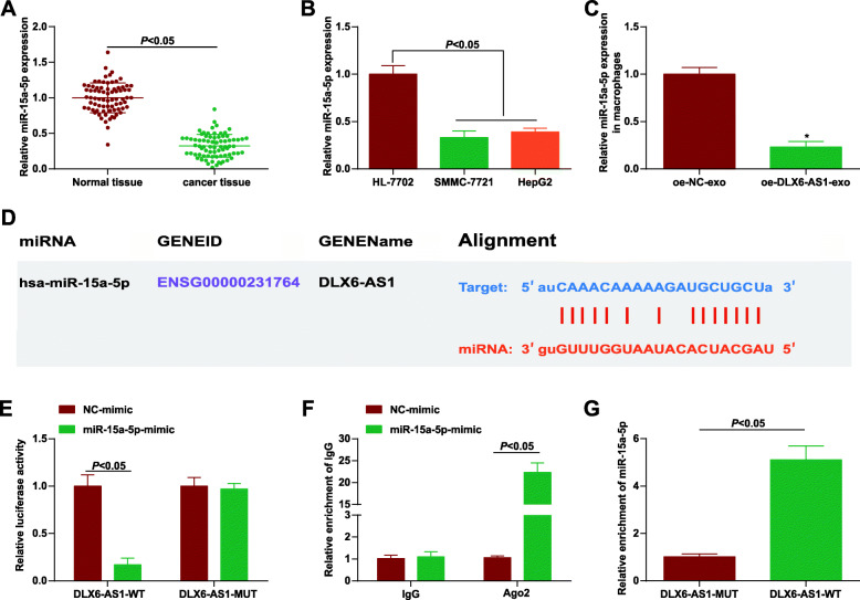
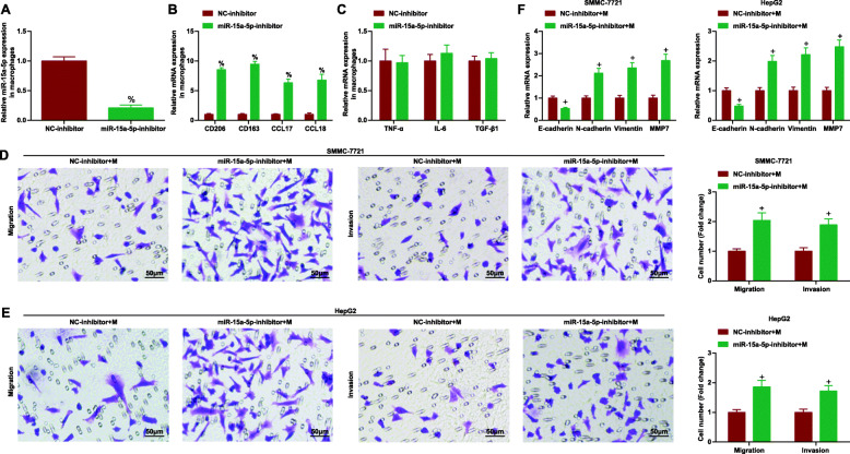
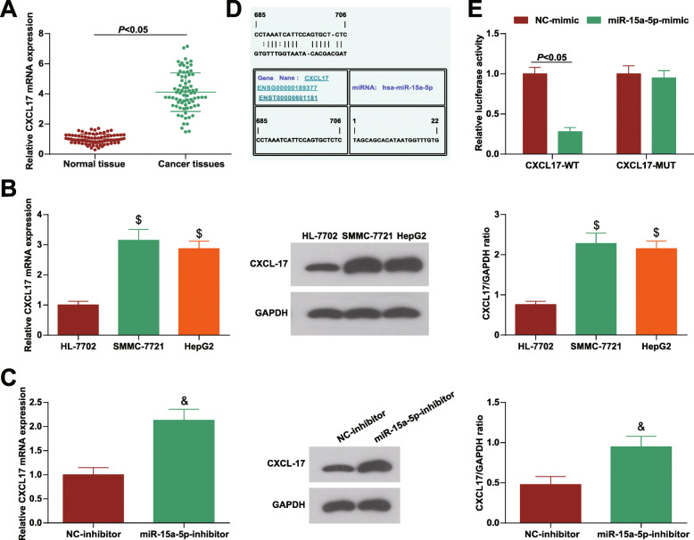
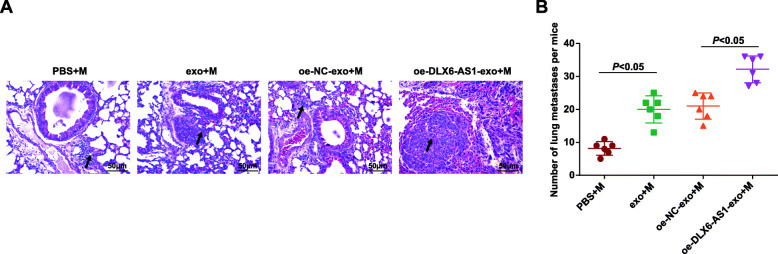
Results: Promoted DLX6-AS1 and CXCL17 and reduced miR-15a-5p exhibited in HCC. HCC-exo induced M2 macrophage polarization to accelerate migration, invasion and epithelial mesenchymal transition in HCC, which was further enhanced by up-regulated DLX6-AS1 but impaired by silenced DLX6-AS1. Inhibition of miR-15a-5p promoted M2 macrophage polarization to stimulate the invasion and metastasis of HCC while that of CXCL17 had the opposite effects. DLX6-AS1 mediated miR-15a-5p to target CXCL17. DLX6-AS1 from HCC-exo promoted metastasis in the lung by inducing M2 macrophage polarization in vivo.

Conclusion: DLX6-AS1 from HCC-exo regulates CXCL17 by competitively binding to miR-15a-5p to induce M2 macrophage polarization, thus promoting HCC migration, invasion and EMT.

Keywords: C-X-C motif chemokine ligand 17; Hepatocellular carcinoma; Hepatocellular carcinoma cell-secreted exosomes; Long non-coding RNA distal-less homeobox 6 antisense 1; M2 macrophages; microRNA-15a-5p.


Conflict of interest statement

The authors declare that they have no conflicts of interest.

Figures

Fig. 1
Fig. 1
HCC-exo induce macrophage M2 polarization. a. Electron microscope observation of the morphology of SMMC-7721 cells-derived exosomes; b. Western blot analysis of antigens (CD63, Alix and TSG101) in SMMC-7721 cells-derived exosomes; c. Microscopic observation of THP1 cells before and after PMA induction; d. PKH26 labeling of exosomes; e. RT-qPCR detection of M2 macrophages markers (CD206, CD163, CCL17 and CCL18) after co-culture with HCC-exo; f. RT-qPCR detection of M1 macrophages markers (TNF-α, IL-6 and TGF-β) after co-culture with HCC-exo; data were expressed as mean ± standard deviation (repetition = 3) and evaluated by One-way AVONA and Tukey’s test. & P < 0.05 compared with the PBS group
Fig. 2
Fig. 2
HCC-exo promote migration, invasion and EMT via inducing M2 macrophage polarization in HCC. a. Transwell assay tested the invasion and migration of SMMC-7721 cells after co-culture with HCC-exo-treated macrophages; b. Transwell assay tested the invasion and migration of HepG2 cells after co-culture with HCC-exo-treated macrophages; c. RT-qPCR analysis of E-cadherin, N-cadherin, vimentin and MMP7 mRNA expression in SMMC-7721 and HepG2 cells after co-culture with HCC-exo-treated macrophages; data were expressed as mean ± standard deviation (repetition = 3) and evaluated by One-way AVONA and Tukey’s test. * P < 0.05 compared with the SMMC-7721 + M group; ^ P < 0.05 compared with the HepG2 + M group
Fig. 3
Fig. 3
DLX6-AS1 is overexpressed and triggers migration, invasion and EMT in HCC. a. RT-qPCR detection of DLX6-AS1 expression in cancer tissues and normal tissues, as well as cancer cells and normal liver cells; b. RT-qPCR detection of DLX6-AS1 expression in SMMC-7721 and HepG2 cells after up-regulating DLX6-AS1; c. Transwell assay tested the invasion and migration of SMMC-7721 cells after up-regulating DLX6-AS1; d. Transwell assay tested the invasion and migration of HepG2 cells after up-regulating DLX6-AS1; e. RT-qPCR analysis of E-cadherin, N-cadherin, vimentin and MMP7 mRNA expression in SMMC-7721 and HepG2 cells after up-regulating DLX6-AS1; data were expressed as mean ± standard deviation (repetition = 3) and evaluated by One-way AVONA and Tukey’s test. $ P < 0.05 compared with the HL-7702 cells; * P < 0.05 compared with the oe-NC group
Fig. 4
Fig. 4
DLX6-AS1 from HCC-exo stimulates M2 macrophage polarization to accelerate migration, invasion and EMT in HCC. a. RT-qPCR detection of DLX6-AS1 expression in serum of HCC patients and healthy controls; b. Detection of CD68 expression in clinical samples through immunohistochemical staining; c. Correlation between expression of DLX6-AS1 and CD68 was analyzed using Pearson test; d. RT-qPCR detection of DLX6-AS1 expression in HCC-exo delivering oe-DLX6-AS1; e. RT-qPCR detection of DLX6-AS1 expression in macrophages after co-culture with HCC-exo delivering oe-DLX6-AS1; f. RT-qPCR detection of M2 macrophages markers (CD206, CD163, CCL17 and CCL18) after co-culture with HCC-exo carrying oe-DLX6-AS1; g. RT-qPCR detection of M1 macrophages markers (TNF-α, IL-6 and TGF-β) after co-culture with HCC-exo carrying oe-DLX6-AS1; h. Transwell assay tested the migration and invasion of SMMC-7721 cells after co-culture with M2 macrophages induced by HCC-exo carrying oe-DLX6-AS1; i. Transwell assay tested the migration and invasion of HepG2 cells after co-culture with M2 macrophages induced by HCC-exo delivering oe-DLX6-AS1; j. RT-qPCR of E-cadherin, N-cadherin, vimentin and MMP7 mRNA expression in SMMC-7721 and HepG2 cells after co-culture with M2 macrophages induced by HCC-exo delivering oe-DLX6-AS1; data were expressed as mean ± standard deviation (repetition = 3) and evaluated by One-way AVONA and Tukey’s test. * P < 0.05 compared with the oe-NC group; # P < 0.05 compared with the PBS group; & P < 0.05 compared with the oe-NC-exo group; ^ P < 0.05 compared with the oe-NC-exo + M group
Fig. 5
Fig. 5
DLX6-AS1 from HCC-exo stimulates M2 macrophage polarization to accelerate migration, invasion and EMT in HCC. a. RT-qPCR detection of DLX6-AS1 expression in HCC-exo delivering sh-DLX6-AS1; b. RT-qPCR detection of DLX6-AS1 expression in macrophages after co-culture with HCC-exo delivering sh-DLX6-AS1; c. RT-qPCR detection of M2 macrophages markers (CD206, CD163, CCL17 and CCL18) after co-culture with HCC-exo carrying sh-DLX6-AS1; d. RT-qPCR detection of M1 macrophages markers (TNF-α, IL-6 and TGF-β) after co-culture with HCC-exo carrying sh-DLX6-AS1; e. Transwell assay tested the migration and invasion of SMMC-7721 cells after co-culture with M2 macrophages induced by HCC-exo carrying sh-DLX6-AS1; f. Transwell assay tested the migration and invasion of HepG2 cells after co-culture with M2 macrophages induced by HCC-exo delivering sh-DLX6-AS1; g/h. RT-qPCR of E-cadherin, N-cadherin, vimentin and MMP7 mRNA expression in SMMC-7721 and HepG2 cells after co-culture with M2 macrophages induced by HCC-exo delivering sh-DLX6-AS1; data were expressed as mean ± standard deviation (repetition = 3) and evaluated by One-way AVONA and Tukey’s test. ^P < 0.05 compared with the sh-NC group; *P < 0.05 compared with the oe-NC-exo group; + P < 0.05 compared with the oe-NC-exo + M group
Fig. 6
Fig. 6
DLX6-AS1 interacts with miR-15a-5p. a. RT-qPCR detection of miR-15a-5p expression in cancer tissues and normal tissues; b. RT-qPCR detection of miR-15a-5p expression in cancer cells and normal liver cells; c. RT-qPCR detection of miR-15a-5p expression in macrophages after co-culture with HCC-exo delivering oe-DLX6-AS1; d. Prediction of the potential binding sites of DLX6-AS1 and miR-15a-5p through bioinformatics websites; e. Verification of the targeting relationship between DLX6-AS1 and miR-15a-5p through dual luciferase reporter experiment; f. RIP detection of the binding relationship between DLX6-AS1 and miR-15a-5p; g. RNA pull-down detection of the binding relationship between DLX6-AS1 and miR-15a-5p; data were expressed as mean ± standard deviation (repetition = 3) and evaluated by One-way AVONA and Tukey’s test. * P < 0.05 compared with the oe-NC-exo group
Fig. 7
Fig. 7
Down-regulating miR-15a-5p promotes M2 macrophage polarization and HCC cell migration, invasion and EMT. a. RT-qPCR detection of miR-15a-5p expression in macrophages after down-regulating miR-15a-5p; b. RT-qPCR detection of M2 macrophages markers (CD206, CD163, CCL17 and CCL18) after down-regulating miR-15a-5p; c. RT-qPCR detection of M1 macrophages markers (TNF-α, IL-6 and TGF-β) after down-regulating miR-15a-5p; d. Transwell assay tested the migration and invasion of SMMC-7721 cells after co-culture with macrophages inhibiting miR-15a-5p; e. Transwell assay tested the migration and invasion of HepG2 cells after co-culture with macrophages inhibiting miR-15a-5p; f. E-cadherin, N-cadherin, vimentin and MMP7 expression in SMMC-7721 and HepG2 cells after co-culture with macrophages inhibiting miR-15a-5p by qPCR; data were expressed as mean ± standard deviation (repetition = 3) and evaluated by One-way AVONA and Tukey’s test. % P < 0.05 compared with the NC-inhibitor group; + P < 0.05 compared with the NC-inhibitor + M group
Fig. 8
Fig. 8
DLX6-AS1 mediates miR-15a-5p to target CXCL17 to drive migration, invasion and EMT in HCC. a. RT-qPCR analysis of CXCL17 expression in clinical tissues; b. RT-qPCR and Western blot analysis of CXCL17 expression in cancer cells and normal liver cells; c. RT-qPCR and Western blot analysis of CXCL17 expression in macrophages after down-regulating miR-15a-5p; d. Prediction of the potential binding site of miR-15a-5p and CXCL17 through bioinformatics websites; e. Verification of the targeting relationship between miR-15a-5p and CXCL17 through dual luciferase reporter experiment; data were expressed as mean ± standard deviation (repetition = 3) and evaluated by One-way AVONA and Tukey’s test. $ P < 0.05 compared with HL-7702 cells; & P < 0.05 compared with the NC-inhibitor group
Fig. 9
Fig. 9
DLX6-AS1 mediates miR-15a-5p to target CXCL17 to drive migration, invasion and EMT in HCC. a. RT-qPCR and Western blot analysis of CXCL17 expression in macrophages after silencing CXCL17; b. RT-qPCR detection of M2 macrophages markers (CD206, CD163, CCL17 and CCL18) after silencing CXCL17; c. RT-qPCR detection of M1 macrophages markers (TNF-α, IL-6 and TGF-β) after silencing CXCL17;d. Transwell assay tested the migration and invasion of SMMC-7721 cells after co-culture with macrophages inhibiting CXCL17; e. Transwell assay tested the migration and invasion of HepG2 cells after co-culture with macrophages inhibiting CXCL17; f. E-cadherin, N-cadherin, vimentin and MMP7 mRNA expression in SMMC-7721 and HepG2 cells after co-culture with macrophages inhibiting CXCL17; data were expressed as mean ± standard deviation (repetition = 3) and evaluated by One-way AVONA and Tukey’s test; % P < 0.05 compared with the si-NC group; + P < 0.05 compared with the si-NC + M group
Fig. 10
Fig. 10
DLX6-AS1 mediates miR-15a-5p to target CXCL17 to drive migration, invasion and EMT in HCC. a. RT-qPCR analysis of CXCL17 expression in macrophages; b. RT-qPCR detection of M2 macrophages markers (CD206, CD163, CCL17 and CCL18); c. RT-qPCR detection of M1 macrophages markers (TNF-α, IL-6 and TGF-β); d. Transwell assay tested the migration and invasion of SMMC-7721 cells; e. Transwell assay tested the migration and invasion of HepG2 cells; f/g. E-cadherin, N-cadherin, vimentin and MMP7 mRNA expression in SMMC-7721 and HepG2 cells; data were expressed as mean ± standard deviation (repetition = 3) and evaluated by One-way AVONA and Tukey’s test. ^ P < 0.05 compared with the oe-DLX6-AS1-exo + si-NC group; + P < 0.05 compared with theoe-DLX6-AS1-exo + si-NC+ M group
Fig. 11
Fig. 11
DLX6-AS1 from HCC-exo promotes lung metastasis by inducing M2 macrophages in mice with HCC. a. H&E-stained tumor tissues (200 ×); b. Number of metastatic hepatic nodules; data were as the mean ± standard deviation (mice = 6/group) and evaluated by One-way AVONA and Tukey’s test

Similar articles

Cited by

References

    1. Vyas M, Zhang X. Hepatocellular carcinoma: role of pathology in the era of precision medicine. Clin Liver Dis. 2020;24(4):591–610. doi: 10.1016/j.cld.2020.07.010. - DOI - PubMed
    1. Roderburg C, Wree A, Demir M, Schmelzle M, Tacke F. The role of the innate immune system in the development and treatment of hepatocellular carcinoma. Hepat Oncol. 2020;7(1):HEP17. doi: 10.2217/hep-2019-0007. - DOI - PMC - PubMed
    1. Trevisan Franca de Lima L, et al. The use of minimally invasive biomarkers for the diagnosis and prognosis of hepatocellular carcinoma. Biochim Biophys Acta Rev Cancer. 2020;1874(2):188451. doi: 10.1016/j.bbcan.2020.188451. - DOI - PubMed
    1. Feng M, et al. Therapy of Primary Liver Cancer. Innovation (N Y) 2020;1(2):100032. - PMC - PubMed
    1. Lopez-Janeiro A, et al. Prognostic value of macrophage polarization markers in epithelial neoplasms and melanoma. A systematic review and meta-analysis. Mod Pathol. 2020;33(8):1458–1465. doi: 10.1038/s41379-020-0534-z. - DOI - PubMed

Publication types

MeSH terms