

Scanxiety: a scoping review about scan-associated anxiety
- PMID: 34039571
- PMCID: PMC8160190
- DOI: 10.1136/bmjopen-2020-043215
Scanxiety: a scoping review about scan-associated anxiety
Abstract
Objectives: To identify available literature on prevalence, severity and contributing factors of scan-associated anxiety ('scanxiety') and interventions to reduce it.
Design: Systematic scoping review.
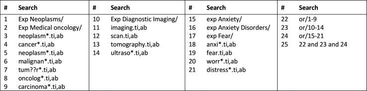
Data sources: Ovid MEDLINE, Ovid EMBASE, Ovid PsycINFO, Ovid Cochrane Central Register of Controlled Trials, Scopus, EBSCO CINAHL and PubMed up to July 2020.
Study selection: Eligible studies recruited people having cancer-related non-invasive scans (including screening) and contained a quantitative assessment of scanxiety.
Data extraction: Demographics and scanxiety outcomes were recorded, and data were summarised by descriptive statistics.
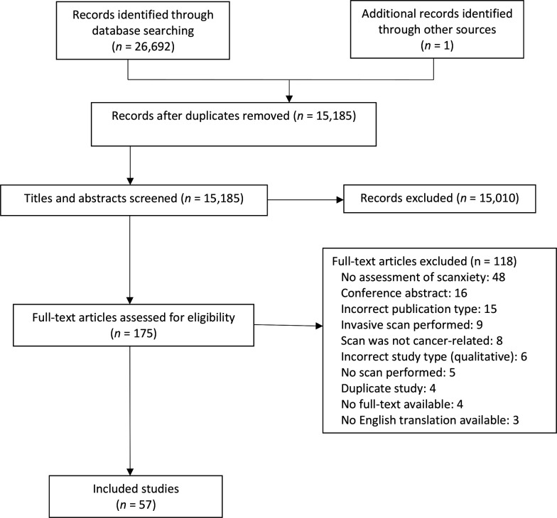
Results: Of 26 693 citations, 57 studies were included across a range of scan types (mammogram: 26/57, 46%; positron-emission tomography: 14/57, 25%; CT: 14/57, 25%) and designs (observation: 47/57, 82%; intervention: 10/57, 18%). Eighty-one measurement tools were used to quantify prevalence and/or severity of scanxiety, including purpose-designed Likert scales (17/81, 21%); the State Trait Anxiety Inventory (14/81, 17%) and the Hospital Anxiety and Depression Scale (9/81, 11%). Scanxiety prevalence ranged from 0% to 64% (above prespecified thresholds) or from 13% to 83% ('any' anxiety, if no threshold). Mean severity scores appeared low in almost all measures that quantitatively measured scanxiety (54/62, 87%), regardless of whether anxiety thresholds were prespecified. Moderate to severe scanxiety occurred in 4%-28% of people in studies using descriptive measures. Nine of 20 studies assessing scanxiety prescan and postscan reported significant postscan reduction in scanxiety. Lower education, smoking, higher levels of pain, higher perceived risk of cancer and diagnostic scans (vs screening scans) consistently correlated with higher scanxiety severity but not age, gender, ethnicity or marital status. Interventions included relaxation, distraction, education and psychological support. Six of 10 interventions showed a reduction in scanxiety.
Conclusions: Prevalence and severity of scanxiety varied widely likely due to heterogeneous methods of measurement. A uniform approach to evaluating scanxiety will improve understanding of the phenomenon and help guide interventions.
Keywords: adult oncology; anxiety disorders; diagnostic radiology.
© Author(s) (or their employer(s)) 2021. Re-use permitted under CC BY-NC. No commercial re-use. See rights and permissions. Published by BMJ.
Conflict of interest statement
Competing interests: None declared.
Figures
Similar articles
-
Prevalence and severity of scanxiety in people with advanced cancers: a multicentre survey.Support Care Cancer. 2022 Jan;30(1):511-519. doi: 10.1007/s00520-021-06454-9. Epub 2021 Aug 1. Support Care Cancer. 2022. PMID: 34333717
-
Telephone interventions for symptom management in adults with cancer.Cochrane Database Syst Rev. 2020 Jun 2;6(6):CD007568. doi: 10.1002/14651858.CD007568.pub2. Cochrane Database Syst Rev. 2020. PMID: 32483832 Free PMC article.
-
Scanxiety among Adults with Cancer: A Scoping Review to Guide Research and Interventions.Cancers (Basel). 2023 Feb 22;15(5):1381. doi: 10.3390/cancers15051381. Cancers (Basel). 2023. PMID: 36900174 Free PMC article.
-
Surveillance-Associated Anxiety After Curative-Intent Cancer Surgery: A Systematic Review.Ann Surg Oncol. 2025 Jan;32(1):47-62. doi: 10.1245/s10434-024-16287-5. Epub 2024 Sep 29. Ann Surg Oncol. 2025. PMID: 39343818 Free PMC article.
-
Experiences with scans and scanxiety in people with advanced cancer: a qualitative study.Support Care Cancer. 2021 Dec;29(12):7441-7449. doi: 10.1007/s00520-021-06319-1. Epub 2021 Jun 2. Support Care Cancer. 2021. PMID: 34076779
Cited by
-
Screening for Brain Metastases in Patients With NSCLC: A Qualitative Study on the Psychologic Impact of Being Diagnosed With Asymptomatic Brain Metastases.JTO Clin Res Rep. 2022 Aug 27;3(10):100401. doi: 10.1016/j.jtocrr.2022.100401. eCollection 2022 Oct. JTO Clin Res Rep. 2022. PMID: 36188631 Free PMC article.
-
Translating radiological research into practice-from discovery to clinical impact.Insights Imaging. 2024 Jan 17;15(1):13. doi: 10.1186/s13244-023-01596-2. Insights Imaging. 2024. PMID: 38228934 Free PMC article.
-
Scan-associated anxiety (scanxiety): the enigma of emotional breathing oscillations at 0.32 Hz (19 bpm).Front Neurosci. 2024 Apr 4;18:1384993. doi: 10.3389/fnins.2024.1384993. eCollection 2024. Front Neurosci. 2024. PMID: 38638691 Free PMC article. Review.
-
Scanxiety and quality of life around follow-up imaging in patients with unruptured intracranial aneurysms: a prospective cohort study.Eur Radiol. 2024 Sep;34(9):6018-6025. doi: 10.1007/s00330-024-10602-0. Epub 2024 Feb 5. Eur Radiol. 2024. PMID: 38311702 Free PMC article.
-
Stress Management Interventions to Facilitate Psychological and Physiological Adaptation and Optimal Health Outcomes in Cancer Patients and Survivors.Annu Rev Psychol. 2023 Jan 18;74:423-455. doi: 10.1146/annurev-psych-030122-124119. Epub 2022 Aug 12. Annu Rev Psychol. 2023. PMID: 35961041 Free PMC article. Review.
References
-
- Feiler B, Scanxiety FB. Scanxiety. fear of a postcancer ritual. Time 2011;177:56. - PubMed
-
- Mathers SA, McKenzie GA, Robertson EM. A necessary evil: the experiences of men with prostate cancer undergoing imaging procedures. Radiography 2011;17:284–91. 10.1016/j.radi.2011.06.005 - DOI
-
- Strand T, Törnqvist E, Rask M, et al. . The experience of patients with neoplasm metastasis in the spine during a magnetic resonance imaging examination. J Radiol Nurs 2014;33:191–8. 10.1016/j.jradnu.2014.09.001 - DOI
Publication types
MeSH terms
LinkOut - more resources
Full Text Sources
Other Literature Sources
Medical