. 2021 Jun 1;118(22):e2102940118.
doi: 10.1073/pnas.2102940118.

Oviductal motile cilia are essential for oocyte pickup but dispensable for sperm and embryo transport

Affiliations

Oviductal motile cilia are essential for oocyte pickup but dispensable for sperm and embryo transport

Shuiqiao Yuan et al. Proc Natl Acad Sci U S A. .

Abstract

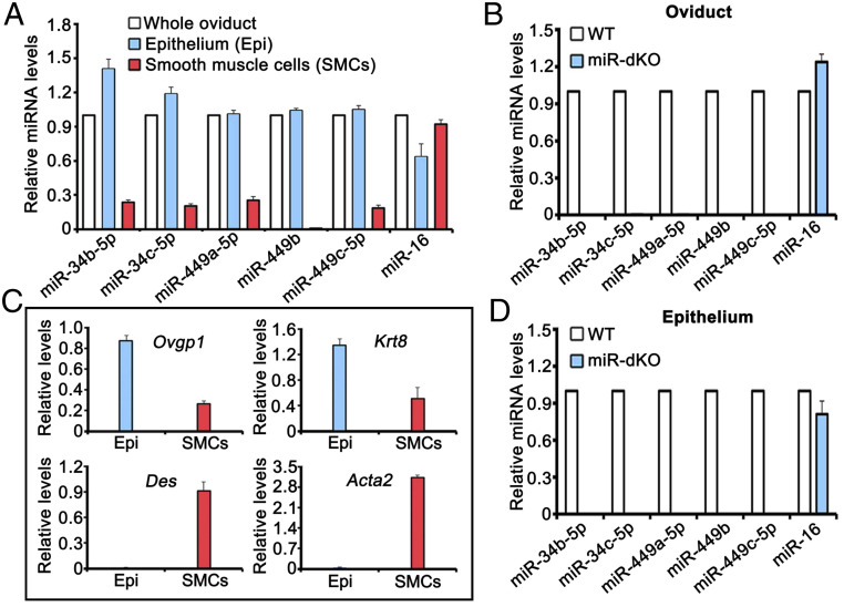
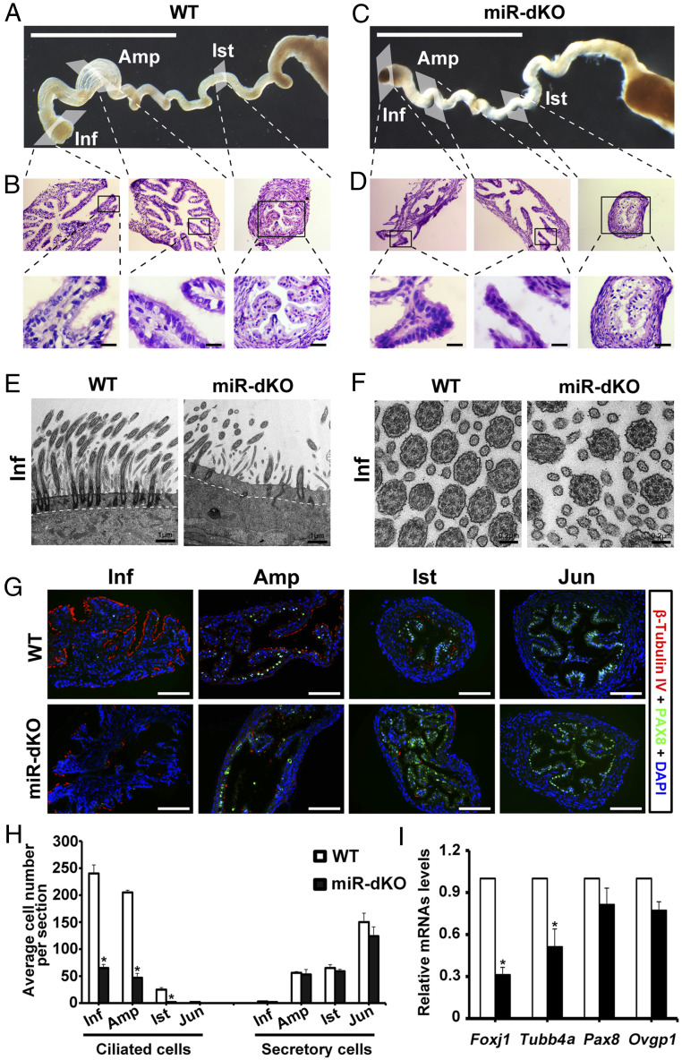
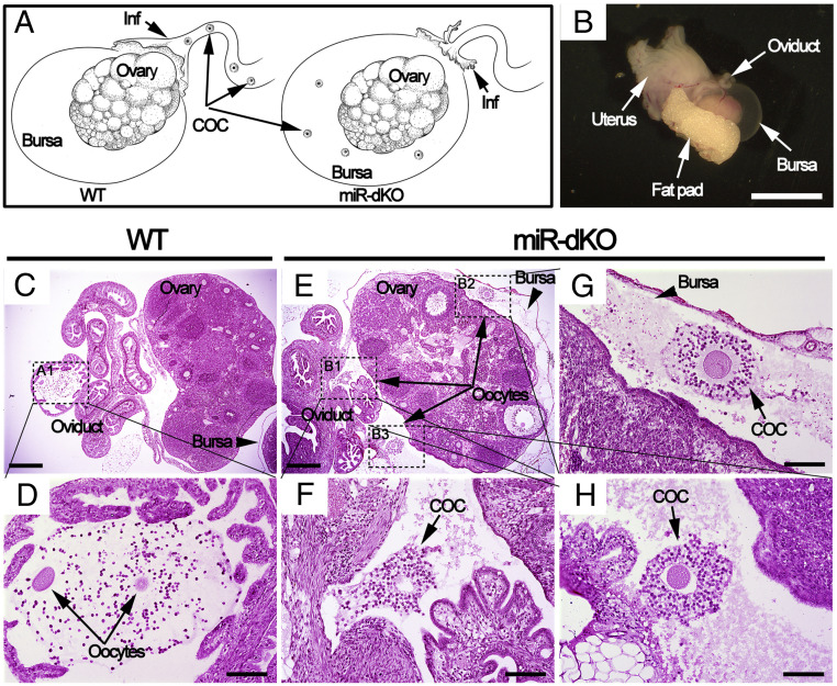
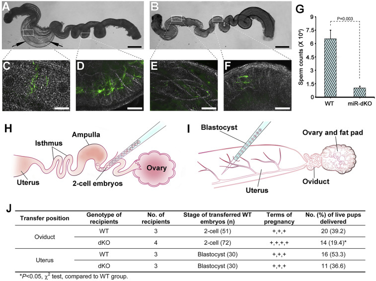
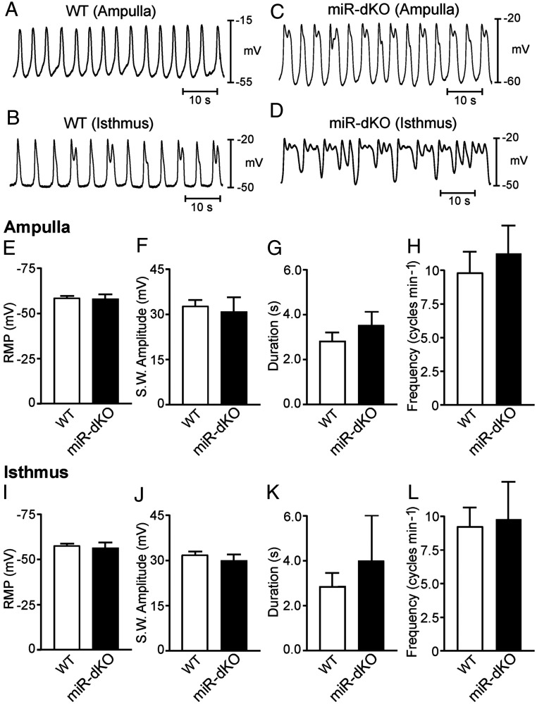
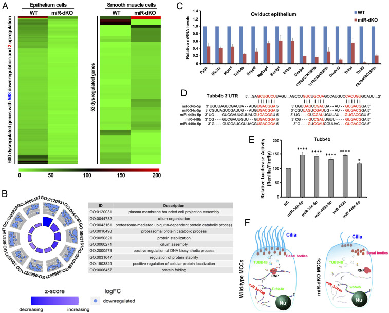
Mammalian oviducts play an essential role in female fertility by picking up ovulated oocytes and transporting and nurturing gametes (sperm/oocytes) and early embryos. However, the relative contributions to these functions from various cell types within the oviduct remain controversial. The oviduct in mice deficient in two microRNA (miRNA) clusters (miR-34b/c and miR-449) lacks cilia, thus allowing us to define the physiological role of oviductal motile cilia. Here, we report that the infundibulum without functional motile cilia failed to pick up the ovulated oocytes. In the absence of functional motile cilia, sperm could still reach the ampulla region, and early embryos managed to migrate to the uterus, but the efficiency was reduced. Further transcriptomic analyses revealed that the five messenger ribonucleic acids (mRNAs) encoded by miR-34b/c and miR-449 function to stabilize a large number of mRNAs involved in cilium organization and assembly and that Tubb4b was one of their target genes. Our data demonstrate that motile cilia in the infundibulum are essential for oocyte pickup and thus, female fertility, whereas motile cilia in other parts of the oviduct facilitate gamete and embryo transport but are not absolutely required for female fertility.

Keywords: Fallopian tube; ciliopathy; fertility; microRNAs; multiciliogenesis.


Conflict of interest statement

The authors declare no competing interest.

Figures

Fig. 1.
Fig. 1.
Five miRNAs encoded by the miR-34b/c and miR-449 clusters are preferentially expressed in the epithelial cells of the oviducts. (A) qPCR analyses of the five miRNAs (miR-34b/c and miR-449a/b/c) in the total oviduct, oviductal epithelial, and smooth muscle cells purified from WT female mice. miR-16 was included as a positive control. (B) qPCR analyses of the five miRNAs (miR-34b/c and miR-449a/b/c) in WT and miR-dKO oviducts. miR-16 was included as a positive control. (C) qPCR analyses of levels of oviductal marker genes, including Ovgp1 and Krt8 (oviductal epithelial cell markers) and Des and Acta2 (smooth muscle cell makers), in enriched epithelial and smooth muscle cells from WT oviducts. (D) qPCR analyses of the five miRNAs (miR-34b/c and miR-449a/b/c) in WT and miR-dKO oviductal epithelial cells. miR-16 was included as a positive control. All qPCR assays were performed in biological triplicates, and the data were presented as mean ± SEM.
Fig. 2.
Fig. 2.
Ablation of miR-34b/c and miR-449 disrupts multiciliogenesis in the oviductal epithelium. (A) Gross morphology of WT oviducts. (Scale bar, 1 mm.) (B) Low-power images showing histology of the WT oviductal infundibulum (Inf), ampulla (Amp), and isthmus (Ist) from the boxed areas in A (Upper). Lower are higher magnifications of the boxed areas from Upper, showing the presence of motile cilia pointing to the lumen. (Scale bar, 10 μm.) (C) Gross morphology of miR-dKO oviducts. (Scale bar, 1 mm.) (D) Low-power images showing histology of the miR-dKO oviductal infundibulum (Inf), ampulla (Amp), and isthmus (Ist) from the boxed areas in C (Upper). Lower are higher magnifications of the boxed areas from Upper, showing the absence of motile cilia. (Scale bar, 10 μm.) (E and F) Ultra-structure of the ciliated epithelium in WT and miR-dKO oviducts revealed by TEM. The miR-dKO epithelium contains much fewer cilia with fewer and misaligned basal bodies, as compared to WT control (E). However, the typical ciliary “9+2” microtubules are present in both WT and miR-dKO controls (F). Scale bars are marked on the images. Five WT and miR-dKO mice were analyzed, and representative images are shown. (G) Representative immunofluorescence images showing β-tubulin IV–positive (ciliated) and PAX8-positive (secretory) cells in infundibulum (Inf), ampulla (Amp), and isthmus (Ist) of the WT and miR-dKO oviducts. (Scale bar, 100 μm.) (H) Quantification of ciliated and secretory cells in Inf, Amp, Ist, and uterotubal junction of WT and miR-dKO oviducts based on immunofluorescent labeling. Data are presented as mean ± SEM (n = 3), *P < 0.05. (I) qPCR analyses of marker genes for ciliated (Foxj1 and Tubb4a) and secretory (Pax8 and Ovgp1) cells in WT and miR-dKO oviducts. Data are presented as mean ± SEM (n = 3), *P < 0.05.
Fig. 3.
Fig. 3.
Ovulated oocytes are trapped inside the ovarian bursa cavity in miR-dKO female mice, as revealed by whole-mount histology analyses. (A) Schematics showing our hypothesis; that is, the ovulated COCs are picked up by fimbriae of the infundibulum and further transported to ampulla for fertilization in WT oviducts (Left), whereas the ovulated COCs fail to be picked up by fimbriae of the infundibulum in miR-dKO female mice and consequently fall into the ovarian bursa cavity (Right). (B) Gross morphology of the female reproductive system ∼3 h after ovulation, including the oviduct, bursa, ovary, fat pad, and uterus. (Scale bar, 5 mm.) (C) A cross-section showing histology of the ovary, oviducts, and bursa. (Scale bar, 0.5 mm.) (D) Digital enlargement of the ampulla region of the oviduct (framed in C), where two oocytes (arrows) are present. (Scale bar, 0.1 mm.) (E) A cross-section showing that the ovulated oocytes are trapped inside the bursa cavity in miR-dKO mice. (Scale bar, 0.5 mm.) (FH) Digital magnification of three areas (framed in E), where individual COCs are present inside the bursa. (Scale bar, 0.1 mm.) Both WT and miR-dKO mice in this experiment were primed with pregnant mare’s serum gonadotropin and hCG, and the samples were collected 15 h after hCG injection. COC, cumulus-oocyte complex; Inf, infundibulum. n = 4 for each genotype.
Fig. 4.
Fig. 4.
The sperm and embryo transport function is compromised in the miR-dKO oviducts. (A and B) Whole oviducts dissected from pregnant mare’s serum gonadotropin/hCG-primed WT (A) and miR-dKO (B) females mated with Stra8-Cre-mTmG+/Tg males. Arrows indicate the presence of oocytes within the oviductal ampulla. (Scale bar, 1 mm.) (C and D) High-power images under transillumination of the two boxed areas in A, showing green sperm inside the oviductal ampulla and isthmus of WT females. (Scale bar, 100 μm.) (E and F) High-power images under transillumination of the two boxed areas in B, showing green sperm inside the oviductal ampulla and isthmus of miR-dKO females. (Scale bar, 100 μm.) (G) Qualification of sperm counts within the oviducts of WT and miR-dKO females ∼3 h after mating with Stra8-Cre-mTmG+/Tg males. Data are presented as mean ± SEM (n = 3). (H and I) Schematics showing transfer of two-cell embryos into WT and miR-dKO oviduct (H), and transfer of blastocyst into WT and miR-dKO uterus (I). (J) Summary of the full-term development of WT embryos transferred into WT and miR-dKO oviducts or uteri.
Fig. 5.
Fig. 5.
Intracellular microelectrode recordings reveal that pacemaker activity in the myosalpinx is comparable between WT and miR-dKO oviducts. (A and B) Representative traces showing spontaneous pacemaker activity termed electrical slow waves recorded from the ampulla (A) and isthmus (B) regions of WT oviducts. (C and D) Representative traces showing slow waves recorded from the ampulla (C) and isthmus (D) regions of miR-dKO oviducts. (EH) Summarized RMP (mV) (E) and slow wave parameters including amplitude (mV) (F), duration (G), and frequency (cycles min−1) (H) in the ampulla region of WT (white bars; n = 6) and miR-dKO (black bars; n = 4) oviducts. (I–L) Summarized RMP (mV) (I) and slow wave parameters including amplitude (mV) (J), duration (K), and frequency (cycles min−1) (L) in the isthmus region of WT (white bars; n = 6) and miR-dKO (black bars; n = 4) oviducts. No statistical differences in membrane potential or slow wave electrical parameters in either ampulla or isthmus regions between WT and miR-dKO oviducts.
Fig. 6.
Fig. 6.
RNA-seq analyses reveal that the five miRNAs encoded by the miR-34b/c and miR-449 clusters control multiciliogenesis by stabilizing their targeted genes in the oviductal epithelium. (A) Heatmap showing significantly dysregulated mRNAs in the oviductal epithelial cells (Left) and smooth muscle cells (Right) of miR-dKO females, as compared to that of WT controls. A total of 600 and 52 dysregulated genes were identified in epithelial cells and smooth muscle cells of miR-dKO oviducts, respectively. (B) Circle plots showing the top 10 Gene Ontology (GO) terms analyzed from 598 down-regulated genes in miR-dKO oviductal epithelial cells. (C) qPCR validation of target gene expression levels in the oviductal epithelium of WT and miR-dKO females. Gapdh was used as an internal control. Data are presented as mean ± SEM (n = 3). (D) A schematic representation showing the two predicted miR-34/449 binding sites in the 3′ UTR of Tubb4b mRNA in mice. (E) Results of luciferase-based reporter assays showing that four out of the five miRNAs (miR-34b/c and miR-449a/b) could stabilize Tubb4b expression. Data are presented as mean ± SEM (n = 3). One-way ANOVA and Bonferroni correction were used for the multiple comparisons. ****P < 0.0001; *P < 0.05. (F) Schematics showing that miR-34b/c and miR-449 clusters regulate multiciliogenesis in oviductal epithelium by stabilizing numerous target genes involved in multiciliogenesis (e.g., Tubb4b). Nu, nucleus; RNP, ribonucleoprotein.

Similar articles

Cited by

References

    1. Stewart C. A., Behringer R. R., “Mouse oviduct development” in Mouse Development, Kubiak J. Z., Ed. (Springer-Verlag, Berlin, Heidelberg, 2012), chap. 14, pp. 247–262. - PubMed
    1. Coy P., García-Vázquez F. A., Visconti P. E., Avilés M., Roles of the oviduct in mammalian fertilization. Reproduction 144, 649–660 (2012). - PMC - PubMed
    1. Halbert S. A., Becker D. R., Szal S. E., Ovum transport in the rat oviductal ampulla in the absence of muscle contractility. Biol. Reprod. 40, 1131–1136 (1989). - PubMed
    1. Halbert S. A., Tam P. Y., Blandau R. J., Egg transport in the rabbit oviduct: The roles of cilia and muscle. Science 191, 1052–1053 (1976). - PubMed
    1. Baker K., Beales P. L., Making sense of cilia in disease: The human ciliopathies. Am. J. Med. Genet. C. Semin. Med. Genet. 151C, 281–295 (2009). - PubMed

Publication types