. 2021 May 26;13(595):eabd7702.
doi: 10.1126/scitranslmed.abd7702.

Transient receptor potential canonical 5 mediates inflammatory mechanical and spontaneous pain in mice

Affiliations

Transient receptor potential canonical 5 mediates inflammatory mechanical and spontaneous pain in mice

Katelyn E Sadler et al. Sci Transl Med. .

Abstract

Tactile and spontaneous pains are poorly managed symptoms of inflammatory and neuropathic injury. Here, we found that transient receptor potential canonical 5 (TRPC5) is a chief contributor to both of these sensations in multiple rodent pain models. Use of TRPC5 knockout mice and inhibitors revealed that TRPC5 selectively contributes to the mechanical hypersensitivity associated with CFA injection, skin incision, chemotherapy induced peripheral neuropathy, sickle cell disease, and migraine, all of which were characterized by elevated concentrations of lysophosphatidylcholine (LPC). Accordingly, exogenous application of LPC induced TRPC5-dependent behavioral mechanical allodynia, neuronal mechanical hypersensitivity, and spontaneous pain in naïve mice. Lastly, we found that 75% of human sensory neurons express TRPC5, the activity of which is directly modulated by LPC. On the basis of these results, TRPC5 inhibitors might effectively treat spontaneous and tactile pain in conditions characterized by elevated LPC.


Conflict of interest statement

Competing interests: The authors have no competing interests to declare with regards to the work contained within this manuscript. T.J.P. and G.D. are cofounders of Ted’s Brain Science, PARMedics, and Doloromics. T.J.P. is also a cofounder of 4E Therapeutics and serves as a consultant for Grunenthal and Synerkine Pharma. G.D. is a consultant for Schedule 1 Therapeutics and Psychogenics.

Figures

Fig. 1.
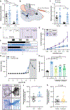
Fig. 1.. TRPC5 contributes to persistent mechanical hypersensitivity in inducible inflammatory pain models.
(A) Depiction of complete Freund’s adjuvant (CFA) model. (B) Mechanical withdrawal thresholds of global TRPC5 knockout (C5KO) and wild-type (WT) mice after CFA injection; n = 11 to 12. (C) Response characterization to noxious needle hindpaw stimulation 9 days after CFA; n = 6 to 10. (D) Response characterization to paintbrush swiping across plantar hindpaw 9 days after CFA; n = 6 to 10. (E) Withdrawal latencies to radiant heat stimulus; n = 10. (F) Mechanical withdrawal thresholds 60 min after intraplantar injection of AC1903; n = 10; arrows indicate injection on each day. (G) Mechanical withdrawal thresholds on day 41 after CFA, 60 min after intraplantar injection of AC1903 (AC) or vehicle (V); n = 11 to 12; Bonferroni post hoc comparison, ***P < 0.001. (H) Mechanical withdrawal thresholds of CFA-injected paw 60 min after intraplantar injection of AC1903 in contralateral paw; n = 5; Bonferroni post hoc comparison, ****P < 0.0001. (I) Depiction of plantar incision model. (J) Mechanical withdrawal thresholds after hindpaw plantar incision; n = 17 to 21. (K) Depiction of spared nerve injury (SNI) and paclitaxel neuropathic pain models. (L) Mechanical withdrawal thresholds after SNI; n = 7 to 10. (M) Mechanical withdrawal thresholds after paclitaxel; n = 8 to 10; arrows indicate paclitaxel injections. Three-way analysis of variance (ANOVA) performed on (B), (E) to (G), (J), (L), and (M); chi-square performed on (C) and (D); two-way ANOVA performed on (H). Post hoc comparisons (Bonferroni or Fisher’s exact) for all panels unless otherwise stated, *P < 0.05, **P < 0.01, and ***P < 0.001 wild-type control versus injury; $P < 0.05 and $ $P < 0.01 C5KO control versus injury; #P < 0.05 and ##P < 0.01 injury C5KO versus wild-type. All data are means ± SEM; B, baseline; AC1903 dose, 50 µg.
Fig. 2.
Fig. 2.. Mouse Trpc5 is expressed in peripheral sensory neurons.
(A) Representative 20× and 40× images of mouse DRG after RNAscope in situ hybridization for Trpc5 (red), CGRP (Calca; green), P2X3R (P2rx3; blue), and NF200 (neurofilament 200; cyan); scale bar, 50 µm (20×) and 20 µm (40×). Arrowheads in 40× image highlight P2rx3-positive neuron and NF200-positive neuron that express Trpc5. (B) Percentage of DRG neurons that express Trpc5; n = 3 mice. (C) Size distribution of Trpc5-positive neurons; n = 1476 neurons from three mice; bars are averages. (D) Coexpression characterization of Trpc5-positive neurons; pie wedges are averages; n = 1476 neurons from three mice.
Fig. 3.
Fig. 3.. Peripherally expressed TRPC5 mediates mechanical hypersensitivity after injuries associated with elevated concentrations of LPC.
(A) Schematic of teased fiber recordings. (B) Representative C fiber activity traces during force ramp application (10 s; up to 100 mN). Force ramps were used to determine the mechanical threshold of each fiber. (C) Mechanical thresholds of fibers from CFA-injected wild-type and C5KO mice; n = 27 to 30 units from five mice; Mann-Whitney test, ***P < 0.001. (D) Representative C fiber activity during static force application. (E) Mean mechanically induced firing rates of C fibers from CFA-injected mice; n = 27 to 30 units from five mice, two-way ANOVA effect of genotype, *P < 0.05. (F) Amount of LPC in skin 2 hours and 7 days after CFA injection; n = 3 to 6. (G) Amount of LPC in skin 1 and 5 days after hindpaw plantar incision; n = 3 to 6. (H) Amount of LPC in sciatic nerve 10 days after SNI; n = 3. (I) Amount of LPC in skin 21 days after first paclitaxel injection; n = 3. Two-way ANOVA performed on (F) to (I). Bonferroni post hoc comparisons for all panels, *P < 0.05, ***P < 0.001, and ****P < 0.0001. All data are means ± SEM.
Fig. 4.
Fig. 4.. LPC induces behavioral and neuronal mechanical hypersensitivity in TRPC5-dependent fashion.
(A) Mechanical withdrawal thresholds of C5KO or wild-type mice 45 to 60 min after intraplantar injection of 50 µM LPC; n = 10. (B) Schematic of dural injection and facial von Frey testing. (C) Periorbital mechanical withdrawal thresholds 60 min after dural application of 4.8 nM LPC and/or 50 µg of AC1903; n = 10. (D) Representative current traces from trigeminal ganglia (TG) neurons during mechanical stimulation. (E) Mean mechanical current density of TG neurons during 250 nM LPC incubation; n = 24 to 30 neurons from three to five mice. (F) Characterization of TG mechanical currents as rapidly adapting (RA), intermediate adapting (IA), slowly adapting (SA), or mechanically insensitive (MI); n =30 to 36 neurons from >3 mice. (G) LPC-mediated calcium flux in HEK cells untransfected or stably expressing mTRPC5; n = 3 independent replicates performed in triplicate. (H) Quantification of LPC (32 µM)–induced calcium flux in HEK cells untransfected or stably expressing mTRPC5 and incubated with ML204 (320 µM), HC-070 (100 µM), or AC1903 (320 µM); n = 3 or more independent replicates performed in triplicate; Bonferroni post hoc, ###P < 0.001 wild-type versus inhibitor. (I) Representative track plots of wild-type and C5KO mice in two-chamber box during postconditioning test; subcutaneous LPC injection (5 µM) associated with purple box, vehicle injection associated with white box. (J) Difference in LPC chamber time between pre- and postconditioning trials; n = 8 to 13; unpaired t test, *P < 0.05. (K) Difference in HC-070 chamber time between pre- and postconditioning trials; n = 18 to 20; unpaired t test, P = 0.06. Two-way ANOVA performed on (A), (C), and (G); chi-square performed on (F); three-way ANOVA performed on (E); one-way ANOVA performed on (H). Bonferroni post hoc comparisons for all panels, *P < 0.05, ***P < 0.001, and ****P < 0.0001. All data are means ± SEM.
Fig. 5.
Fig. 5.. After initial exposure to LPC, TRPC5 activity mediates mechanical hypersensitivity even in the absence of LPC.
(A) Mean mechanical current density of TG neurons isolated from mice 24 hours after dural application of 4.8 nM LPC; n = 33 to 45 cells from three to five mice; three-way ANOVA, followed by Bonferroni post hoc comparison, **P < 0.01 wild-type vehicle versus LPC. (B) Periorbital mechanical withdrawal thresholds after dural application of 1.2 nM LPC and pH 7.0 PBS; n = 10 to 15; purple arrow indicates LPC injection, yellow arrow indicates pH 7.0 PBS injection; three-way ANOVA, followed by Bonferroni post hoc comparison, **P < 0.01 wild-type vehicle versus LPC and ## P < 0.01 LPC wild-type versus C5KO. (C) Relative amount of LPC in serum of wild-type and sickle cell disease (SCD) mice; n = 3, unpaired t test *P < 0.05. (D) Mechanical withdrawal thresholds 60 min after intraplantar injection of AC1903 (50 µg); n = 10 to 14; two-way ANOVA, followed by Bonferroni post hoc comparisons, *P < 0.05. (E) Response characterization to noxious needle hindpaw stimulation 60 min after intraplantar injection of AC1903 (50 µg); n = 9 to 10; chi-square, followed by Fisher’s exact test, **P < 0.01 vehicle wild-type versus SCD, #P < 0.05, and ##P < 0.01 SCD vehicle versus AC1903. (F) Characterization of DRG mechanical currents as RA, IA, SA, or MI; chi-square, followed by Fisher’s exact test, #P < 0.05 and ##P < 0.01 SCD vehicle versus AC1903; n = 28 to 32 neurons from three to five mice. All data are means ± SEM unless otherwise stated.
Fig. 6.
Fig. 6.. hTRPC5 is expressed by most of the DRG neurons and activated by LPC.
(A) Representative 20× image of human DRG neurons after RNAscope in situ hybridization for TRPC5 (red), CGRP (CALCA; green), P2XR3 (P2RX3; blue), and DAPI (magenta); scale bar, 50 µM. (B) Percentage of DRG neurons that express TRPC5; n = 3 humans. (C) Frequency of TRPC5-positive neurons across population of human DRG neurons; n = 438 neurons from three humans; bars are averages. (D) Coexpression characterization of TRPC5-positive and TRPC5-negative neurons; pie wedges are averages; n = 580 neurons from three humans. (E) LPC-mediated calcium flux in HEK cells untransfected or stably expressing hTRPC5; n = 3 independent replicates performed in triplicate. (F) Quantification of LPC (32 µM)–induced calcium flux in HEK cells untransfected or stably expressing hTRPC5 and incubated with ML204 (320 µM), HC-070 (100 µM), or AC 1903 (320 µM); n = 3 independent replicates performed in triplicate; two-way ANOVA performed on (E), one-way ANOVA performed on (F), Bonferroni post hoc comparisons, *P < 0.05 and ***P < 0.001. All data are means ± SEM unless otherwise stated.

Comment in

Similar articles

Cited by

References

    1. Lolignier S, Eijkelkamp N, Wood JN, Mechanical allodynia. Pflugers Arch 467, 133–139 (2014). - PMC - PubMed
    1. Woolf CJ, Central sensitization: Implications for the diagnosis and treatment of pain. Pain 152, S2–S15 (2011). - PMC - PubMed
    1. Eid SR, Crown ED, Moore EL, Liang HA, Choong KC, Dima S, Henze DA, Kane SA, Urban MO, HC-030031, a TRPA1 selective antagonist, attenuates inflammatory- and neuropathy-induced mechanical hypersensitivity. Mol. Pain 4, 48 (2008). - PMC - PubMed
    1. Szczot M, Liljencrantz J, Ghitani N, Barik A, Lam R, Thompson JH, Bharucha-Goebel D, Saade D, Necaise A, Donkervoort S, Foley AR, Gordon T, Case L, Bushnell MC, Bönnemann CG, Chesler AT, PIEZO2 mediates injury-induced tactile pain in mice and humans. Sci. Transl. Med 10, eaat9892 (2018). - PMC - PubMed
    1. Murthy SE, Loud MC, Daou I, Marshall KL, Schwaller F, Kühnemund J, Francisco AG, Keenan WT, Dubin AE, Lewin GR, Patapoutian A, The mechanosensitive ion channel Piezo2 mediates sensitivity to mechanical pain in mice. Sci. Transl. Med 10, eaat9897 (2018). - PMC - PubMed

Publication types