Review
. 2021 May 26;21(4):292-299.
doi: 10.1136/practneurol-2020-002838. Online ahead of print.

Management of pineal and colloid cysts

Affiliations
Review

Management of pineal and colloid cysts

Michael D Jenkinson et al. Pract Neurol. .

Abstract

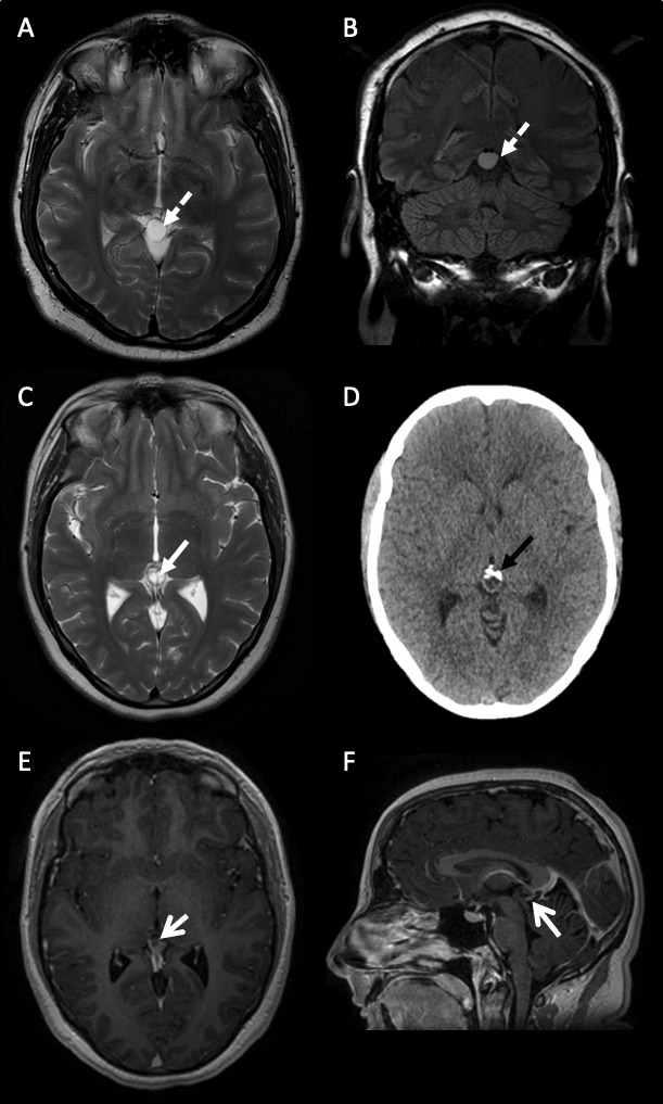
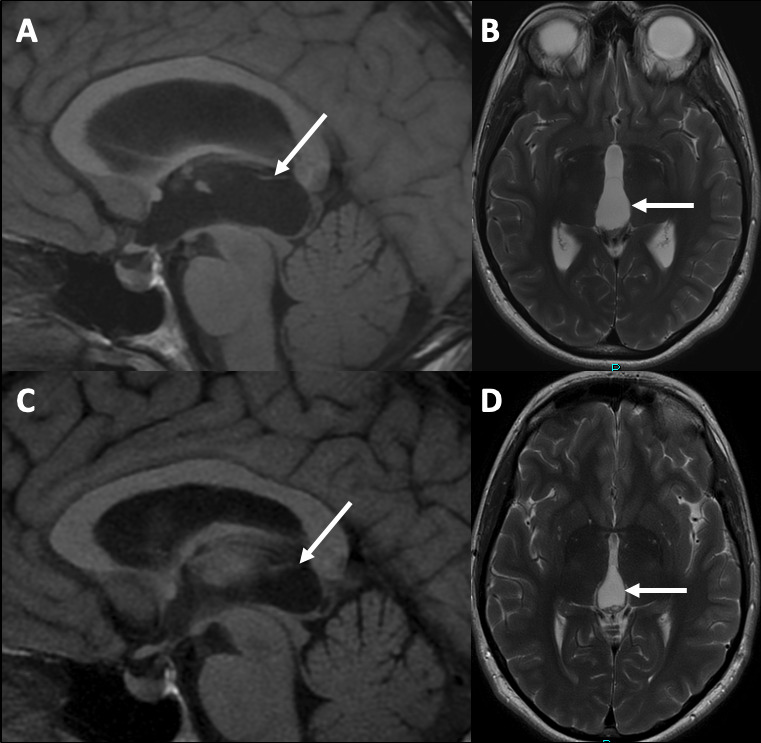
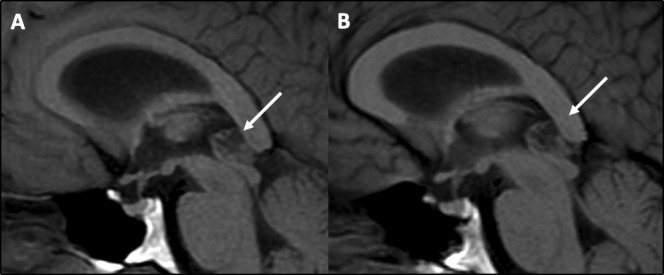
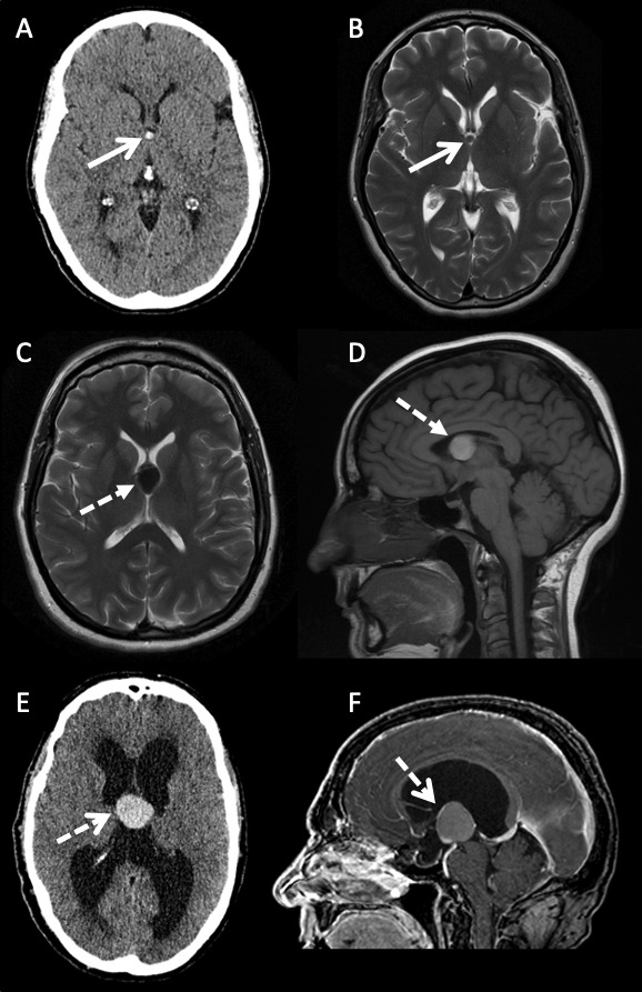
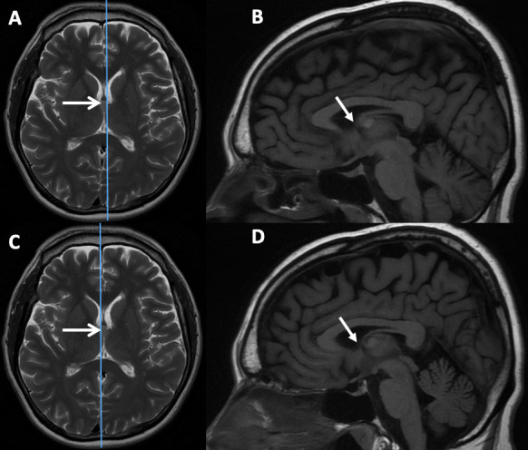
The widespread use of MRI has led to the increasingly frequent diagnosis of pineal and colloid cysts. While most are small and incidental, do not require long-term monitoring and will never need treatment, they are a cause of patient anxiety and clinician uncertainty regarding the optimal management-particularly for larger cysts or those with an atypical appearance. Occasionally pineal cysts, and more commonly colloid cysts, cause hydrocephalus that requires urgent neurosurgical treatment. More recently the non-hydrocephalic symptomatic pineal cyst has been described in the neurosurgical literature but there is controversy over this entity and its management. This review addresses the difficulties in managing pineal and colloid cysts and provides a pragmatic framework for the practising clinician.

Keywords: CSF; clinical neurology; headache; neuroradiology; neurosurgery.


Conflict of interest statement

Competing interests: None declared.

Figures

Figure 1
Figure 1
Varying MR brain scan features of pineal region cysts. (A) and (B) A 38-year-old man with a simple pineal region cyst (A) axial T2-weighted and (B) coronal FLAIR sequences of a simple unilocular pineal region cyst with no internal structure (dashed white arrow). The signal hyperintensity does not suppress on FLAIR imaging. (C–F) A 31-year-old woman with an atypical pineal region cyst. (C) Axial T2-weighted sequence of an atypical multilocular, septated (white closed arrow) pineal region cyst. (D) Axial CT scan of head showing anterior calcification (black closed arrow). (E) Axial and (F) sagittal gadolinium-enhanced postcontrast T1 sequences of an atypical septated, posteriorly enhancing (open white arrows) pineal region cyst. FLAIR (Fluid Attenuated Inversion Recovery).
Figure 2
Figure 2
(A) Sagittal T1-weighted and (B) axial T2-weighted MRI sequences showing a large simple pineal region cyst (white arrows) causing symptomatic hydrocephalus at presentation. (C) Sagittal T1-weighted and (D) axial T2-weighted MRI sequences following endoscopic third ventriculostomy and cyst fenestration with collapse of the pineal region cyst and resolution of the hydrocephalus.
Figure 3
Figure 3
MR scan of brain showing no change in size, shape or appearance on sagittal T1-weighted MRI sequences of an atypical pineal cyst (arrows) in 2016 (A) and 2020 (B).
Figure 4
Figure 4
Varying imaging features of colloid cysts. (A) Axial CT scan and (B) axial T2-weighted sequence MRI of a 46-year-old woman with a small CT hyperattenuating (open arrow) colloid cyst within the anterior aspect of the third ventricle. The signal intensity is intermediate to isointense to white matter on the T2-weighted sequence (open arrows). (C) Axial T2-weighted and (D) sagittal T1-weighted MRI sequences of a 50-year-old woman with a large colloid cyst. The lesion is hypointense on T2 and hyperintense on T1 (dashed closed arrow). There is no hydrocephalus. (E) Axial CT and (F) sagittal postgadolinium contrast T1-weighted MRI sequence of a large colloid cyst in a 59-year-old patient presenting with hydrocephalus. The lesion is hyperattenuating on CT, hyperintense on T1 with some peripheral enhancement secondary to displaced and stretched septal veins (dashed open arrows).
Figure 5
Figure 5
Axial T2-weighted and sagittal T1-weighted MRI sequences showing a colloid cyst (open arrows) arising from the middle part of the third ventricle. The parasagittal cuts to the right (B) and left (D) of midline show that the foramen of Monro is not obstructed (closed arrows) by the colloid cyst, despite the appearance on the T2 axial, therefore the risk of developing symptomatic hydrocephalus requiring treatment is low. The patient is asymptomatic and has been followed for 4 years with no change in MR appearance or clinical features.

References

    1. Tapp E, Huxley M. The histological appearance of the human pineal gland from puberty to old age. J Pathol 1972;108:137–44. 10.1002/path.1711080207 - DOI - PubMed
    1. Sawamura Y, Ikeda J, Ozawa M, et al. . Magnetic resonance images reveal a high incidence of asymptomatic pineal cysts in young women. Neurosurgery 1995;37:11–16. 10.1227/00006123-199507000-00002 - DOI - PubMed
    1. Gokce E, Beyhan M. Evaluation of pineal cysts with magnetic resonance imaging. World J Radiol 2018;10:65–77. 10.4329/wjr.v10.i7.65 - DOI - PMC - PubMed
    1. Nevins EJ, Das K, Bhojak M, et al. . Incidental pineal cysts: is surveillance necessary? World Neurosurg 2016;90:96–102. 10.1016/j.wneu.2016.02.092 - DOI - PubMed
    1. Pu Y, Mahankali S, Hou J, et al. . High prevalence of pineal cysts in healthy adults demonstrated by high-resolution, noncontrast brain MR imaging. AJNR Am J Neuroradiol 2007;28:1706–9. 10.3174/ajnr.A0656 - DOI - PMC - PubMed