

Ndufs4 ablation decreases synaptophysin expression in hippocampus
- PMID: 34040028
- PMCID: PMC8155116
- DOI: 10.1038/s41598-021-90127-4
Ndufs4 ablation decreases synaptophysin expression in hippocampus
Abstract
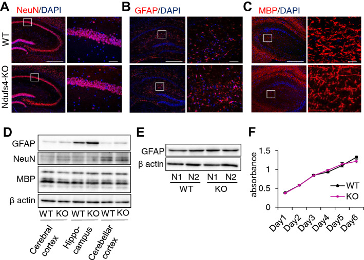
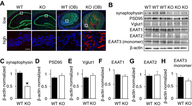
Altered function of mitochondrial respiratory chain in brain cells is related to many neurodegenerative diseases. NADH Dehydrogenase (Ubiquinone) Fe-S protein 4 (Ndufs4) is one of the subunits of mitochondrial complex I and its mutation in human is associated with Leigh syndrome. However, the molecular biological role of Ndufs4 in neuronal function is poorly understood. In this study, upon Ndufs4 expression confirmation in NeuN-positive neurons, and GFAP-positive astrocytes in WT mouse hippocampus, we found significant decrease of mitochondrial respiration in Ndufs4-KO mouse hippocampus. Although there was no change in the number of NeuN positive neurons in Ndufs4-KO hippocampus, the expression of synaptophysin, a presynaptic protein, was significantly decreased. To investigate the detailed mechanism, we silenced Ndufs4 in Neuro-2a cells and we observed shorter neurite lengths with decreased expression of synaptophysin. Furthermore, western blot analysis for phosphorylated extracellular regulated kinase (pERK) revealed that Ndufs4 silencing decreases the activity of ERK signalling. These results suggest that Ndufs4-modulated mitochondrial activity may be involved in neuroplasticity via regulating synaptophysin expression.
Conflict of interest statement
The authors declare no competing interests.
Figures


Similar articles
-
Mitochondrial Function in Astrocytes Is Essential for Normal Emergence from Anesthesia in Mice.Anesthesiology. 2019 Mar;130(3):423-434. doi: 10.1097/ALN.0000000000002528. Anesthesiology. 2019. PMID: 30707122 Free PMC article.
-
Proteomic and metabolomic analyses of mitochondrial complex I-deficient mouse model generated by spontaneous B2 short interspersed nuclear element (SINE) insertion into NADH dehydrogenase (ubiquinone) Fe-S protein 4 (Ndufs4) gene.J Biol Chem. 2012 Jun 8;287(24):20652-63. doi: 10.1074/jbc.M111.327601. Epub 2012 Apr 25. J Biol Chem. 2012. PMID: 22535952 Free PMC article.
-
Mitochondrial dysfunction in GnRH neurons impaired GnRH production.Biochem Biophys Res Commun. 2020 Sep 10;530(1):329-335. doi: 10.1016/j.bbrc.2020.07.090. Epub 2020 Aug 7. Biochem Biophys Res Commun. 2020. PMID: 32828307
-
Ndufs4 knockout mouse models of Leigh syndrome: pathophysiology and intervention.Brain. 2022 Mar 29;145(1):45-63. doi: 10.1093/brain/awab426. Brain. 2022. PMID: 34849584 Free PMC article. Review.
-
Cellular and animal models for mitochondrial complex I deficiency: a focus on the NDUFS4 subunit.IUBMB Life. 2013 Mar;65(3):202-8. doi: 10.1002/iub.1127. Epub 2013 Feb 3. IUBMB Life. 2013. PMID: 23378164 Review.
Cited by
-
Microglial response promotes neurodegeneration in the Ndufs4 KO mouse model of Leigh syndrome.Glia. 2022 Nov;70(11):2032-2044. doi: 10.1002/glia.24234. Epub 2022 Jun 30. Glia. 2022. PMID: 35770802 Free PMC article.
-
Mitochondrial complex I deficiency induces Alzheimer's disease-like signatures that are reversible by targeted therapy.Alzheimers Dement. 2025 Aug;21(8):e70519. doi: 10.1002/alz.70519. Alzheimers Dement. 2025. PMID: 40731203 Free PMC article.
-
GIRUS-net: A Multimodal Deep Learning Model Identifying Imaging and Genetic Biomarkers Linked to Alzheimer's Disease Severity.Annu Int Conf IEEE Eng Med Biol Soc. 2023 Jul;2023:1-4. doi: 10.1109/EMBC40787.2023.10341000. Annu Int Conf IEEE Eng Med Biol Soc. 2023. PMID: 38083359 Free PMC article.
-
Tempo and mode of gene expression evolution in the brain across primates.Elife. 2024 Jan 26;13:e70276. doi: 10.7554/eLife.70276. Elife. 2024. PMID: 38275218 Free PMC article.
References
Publication types
MeSH terms
Substances
LinkOut - more resources
Full Text Sources
Other Literature Sources
Molecular Biology Databases
Research Materials
Miscellaneous
