. 2021 May 26;11(1):11046.
doi: 10.1038/s41598-021-90348-7.

Novel human neutralizing mAbs specific for Spike-RBD of SARS-CoV-2

Affiliations

Novel human neutralizing mAbs specific for Spike-RBD of SARS-CoV-2

Margherita Passariello et al. Sci Rep. .

Abstract

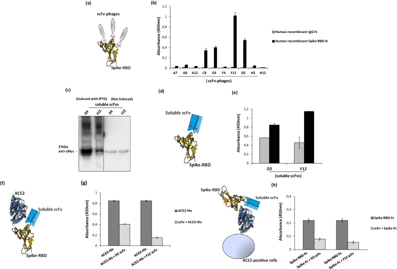
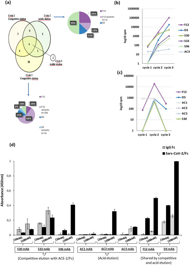
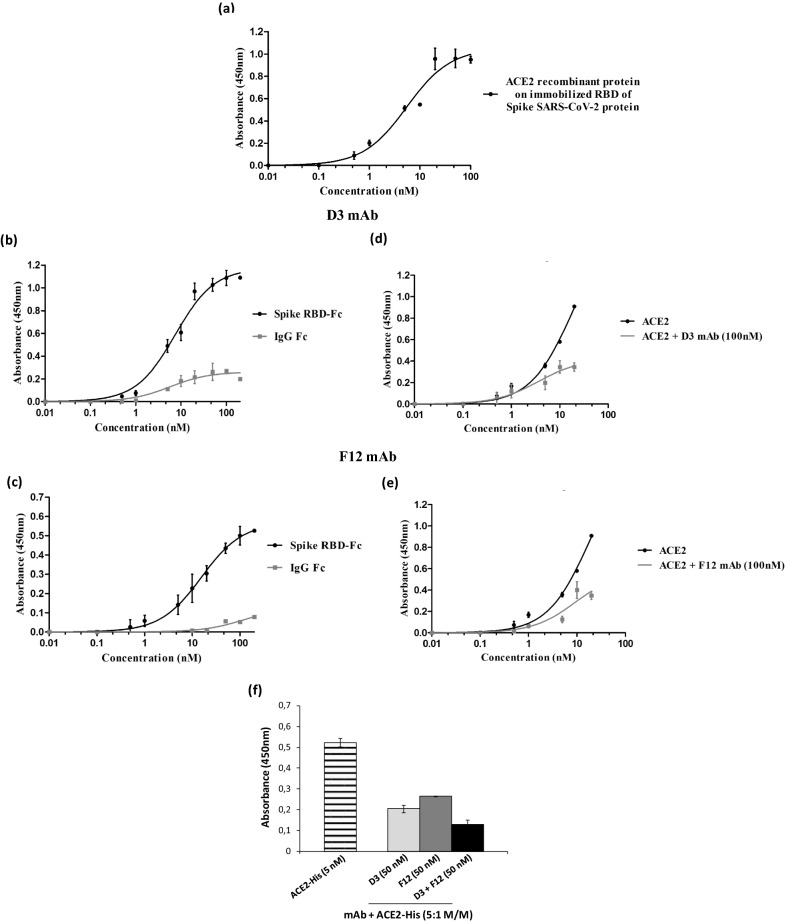
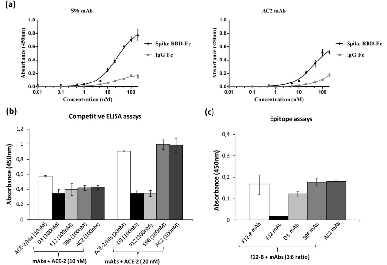
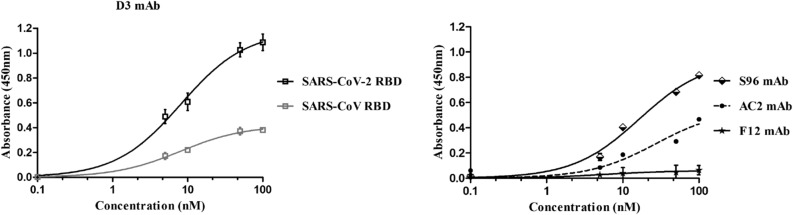
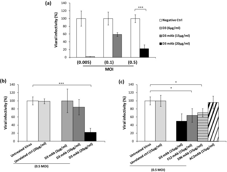
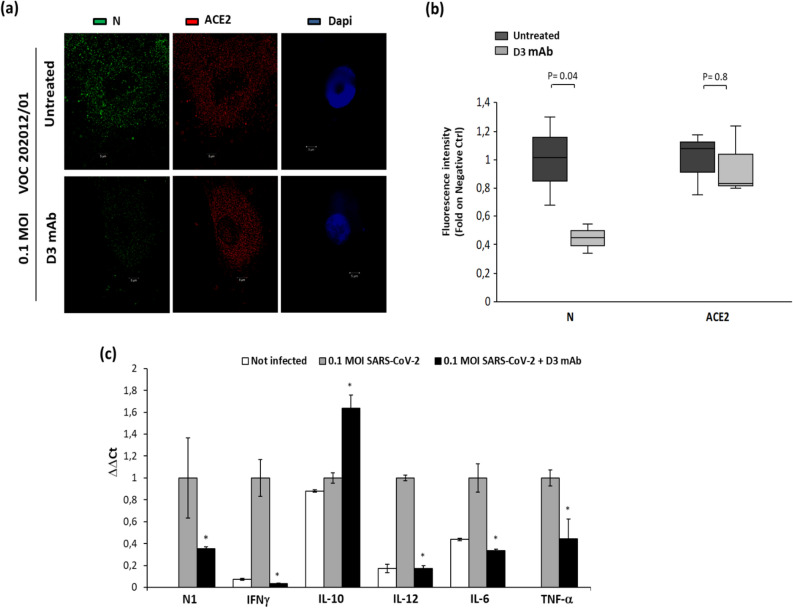
Among the therapies against the pandemic SARS-CoV-2 virus, monoclonal Antibodies (mAbs) targeting the Spike glycoprotein represent good candidates to interfere in the Spike/ACE2 interaction, preventing virus cell entry. Since anti-spike mAbs, used individually, might be unable to block the virus entry in the case of resistant mutations, we designed an innovative strategy for the isolation of multiple novel human scFvs specific for the binding domain (RBD) of Spike. By panning a large phage display antibody library on immobilized RBD, we obtained specific binders by eluting with ACE2 in order to identify those scFvs recognizing the epitope of Spike interacting with its receptor. We converted the novel scFvs into full size IgG4, differently from the previously isolated IgG1 mAbs, to avoid unwanted potential side effects of IgG1 potent effector functions on immune system. The novel antibodies specifically bind to RBD in a nanomolar range and interfere in the interaction of Spike with ACE2 receptor, either used as purified protein or when expressed on cells in its native conformation. Furthermore, some of them have neutralizing activity for virus infection in cell cultures by using two different SARS-CoV-2 isolates including the highly contagious VOC 202012/01 variant and could become useful therapeutic tools to fight against the SARS-CoV-2 virus.


Conflict of interest statement

The authors declare that a patent relative to some of the novel antibodies, mentioned in the manuscript, has been recently filed.

Figures

Figure 1
Figure 1
Schematic representation of selection and screening strategies for identification of SARS-CoV-2 neutralizing mAbs. (a) Cartoon representing SARS-CoV-2 host cell attachment mediated via ACE2-Spike interaction. The ACE2-RBD interaction in the box is adapted from PDB 6M0J. (b) Phage display of scFv library was carried out on recombinant SARS-CoV-2 RBD-Fc protein; acidic or competitive elution were implemented. (c) Variable heavy chains of scFvs were extracted from sub-libraries and sequenced by MiSeq Illumina. (d) The trend of enrichment of given clones was evaluated within and between the selection cycles. (e) Potential binders (scFvs) were converted into fully human IgG4 mAbs in a high yield expressing cell line (HEK293ES_1). (f) Binding to rRBD and competition with rACE2 were evaluated in vitro with recombinant protein assay. Neutralization capacity of RBD-specific mAbs was further evaluated for blocking SARS-CoV-2 replication.
Figure 2
Figure 2
Screening by ELISA and expression of anti-Spike scFvs for analysis of their binding to Spike RBD and their competition with ACE2. (a) Representative image of scFv-phages screening by ELISA to test their binding to Spike-RBD recombinant protein. (b) Screening of positive phage clones by ELISA assay on human Spike RBD-Fc recombinant protein (black bars) or human recombinant IgG Fc used as a negative control in parallel assays (grey bars). (c) Western blotting analysis with the anti-c-myc antibody of the periplasmic extracts of the cells transformed with the two selected positive clones, D3 and F12, expressed in the absence or in the presence of IPTG, used for induction (The blot was obtained by grouping two different parts of the same blot and the black line has been inserted to indicate the two distinct parts. The corresponding full-length blot has been inserted in Supplementary Data set as full-length blot of Figure 2. The samples were processed in parallel in the same experiment). (d) Representative image of soluble scFvs binding to Spike-RBD recombinant protein by ELISA (e) at two different concentrations: 45 nM (grey bars) or 90 nM (black bars). (f) Representative image of soluble scFvs interference in Spike-ACE2 interaction tested by ELISA (g). The binding of ACE2-His to immobilized Spike RBD protein in the absence (dark grey bars) or in the presence (light grey bars) of the indicated soluble scFvs (90 nM). (h) Competitive ELISA assay was performed by measuring the binding of Spike RBD-Fc protein on ACE2-positive VERO E6 cells in the absence or in the presence of D3 and F12 scFvs.
Figure 3
Figure 3
In silico analysis of sub-libraries and identification of potential binders. (a) Venn diagram representing the overlapping of full lenght scFvs among top 100 enriched sequences (in-frame and non-Stop codon bearing) between selection cycles from different elution methods (i.e. acidic and competitive). Cycle 3 from acidic elution was represented apart as only two clones resulted enriched. Pie charts represent the distribution of clones within cycle 2 from acidic elution and cycle 3 from competitive one. F12 (dark violet) and D3 (dark blue) resulted as the most enriched ones. Sequences differing from F12 and D3 in only 1 amino acid were indicated as “variants” (light violet and light blue). (b,c) The enrichment of valid clones between the three selection cycles for competitive elution (b) and acidic elution (c). (d) ELISA assay on human Spike RBD of the converted mAbs: the eight converted mAbs were tested on human Spike RBD-Fc recombinant protein (black bars) or on human Fc (grey bars), used as negative control in parallel assays.
Figure 4
Figure 4
Binding affinity of D3 and F12 mAbs for Spike RBD and their competition with ACE2. (a) The binding affinity of ACE2 receptor to immobilized SARS-CoV-2 RBD recombinant protein was tested by ELISA as a positive control. (b,c) ELISA assays were performed by testing D3 (b) and F12 (c) mAbs at increasing concentrations (0.5–100 nM) on human Spike RBD-Fc chimeric protein (black curves). In parallel the Fc domain (grey curves) was used as a negative control. (d,e) Competitive ELISA assays were performed by measuring the binding of ACE2-His protein to Spike RBD in the absence or in the presence of D3 (d) and F12 (e) mAb used at a concentration of 100 nM. (f) Competitive ELISA assays were performed by measuring the binding of ACE2-His to RBD in the absence (striped bar) or in the presence of the indicated mAbs used alone (gray bars) or in combination (black bars) at a concentration of 100 nM. The binding values were reported as the mean of at least three determinations obtained in three independent experiments. Error bars depicted means ± SD.
Figure 5
Figure 5
ELISA assays on Spike RBD to test the binding affinity of S96 and AC2 mAbs and their competition with ACE-2. (a) The binding affinity and specificity for Spike RBD of S96 and AC2 mAbs was evaluated by testing each mAb at increasing concentrations (0.5–100 nM) on Spike RBD-Fc chimeric protein or Fc, used as a negative control. (b) Competitive ELISA assays were performed by measuring the binding of ACE2-His to RBD in the absence (white bars) or in the presence of the indicated mAbs (gray and black bars) at a concentration of 100 nM. (c) Competitive ELISA assays to determine the epitope binning were performed by measuring the binding of Biotinylated F12 (F12-B) mAb to RBD, pre-incubated in the absence (white bars) or in the presence of the indicated mAbs (grey and black bars). The binding values were reported as the mean of at least three determinations obtained in three independent experiments. Error bars depicted means ± SD.
Figure 6
Figure 6
Cross-reactivity of the novel mAbs for SARS-CoV RBD recombinant protein. The binding of D3 (squares, grey curve), S96 (rhomboids, black curve), F12 (stars, black curve) or AC2 (full circles, black curve) mAbs, tested at increasing concentrations (5–100 nM) on immobilized SARS-CoV RBD protein was analyzed in comparison with the binding of D3 to immobilized SARS-CoV-2 RBD protein (empty squares, black curve on the left) by ELISA assays. Both the RBD proteins were coated on multi-well plates at the concentration of 5 μg/ml and treated as described in “Methods” section. The binding values were reported as the mean of at least three determinations obtained in three independent experiments. Error bars depicted means ± SD.
Figure 7
Figure 7
Neutralization assays to test the biological effects of anti-Spike mAbs. VERO E6 cells were infected with the SARS-CoV-2 virus at the indicated MOI, preincubated in the absence or presence of each mAb used at the indicated concentrations. (a) Viral cell entry and infectivity was measured by RT-PCR of the cell extracts after washes by analyzing the expression of N1 viral gene. The values are expressed as percentage with respect to the negative untreated control. (b) Dose dependent effects of D3 mAb on viral infectivity were measured at the highest viral load. (c) Comparison of the efficiency of the different mAbs for inhibiting virus infectivity of cell cultures at a concentration of 15 µg/ml.
Figure 8
Figure 8
Analysis of human primary cells, infected with the VOC 202012/01 lineage B.1.1.7 variant in the absence or presence of D3, by immunofluorescence assays and RT-PCR. (a) The cells untreated or infected with the VOC 202012/01 variant of SARS-CoV-2, in the absence or presence of D3 for 72 h at 37 °C, were fixed, washed and permeabilised, as described in the Methods. After blocking, the slides were incubated with the relevant primary antibodies overnight at 4 °C: anti-ACE2 (1:100; ab15348; Abcam) or anti-SARS-CoV-2 Nucleoprotein (N) Antibody (1:100; No. 35-579, ProSci), followed by the relevant secondary anti-mouse Alexa Fluor 488 (1:200; ab150113; Abcam) and anti-Rabbit Alexa Fluor 546 (1:200; A-11035; ThermoFisher), respectively. DNA was stained with DAPI (1:5000; #62254; Thermo Fisher). Microscopy images were obtained with the Elyra 7 platform (Zeiss) with the optical Lattice SIM technology, using the 63 × oil immersion objective. (b) Quantification of the mean fluorescence intensity was performed by using the ZEN software (Zeiss, black edition). (c) Cell extracts were analyzed by RT-PCR for measuring the expression of N1 viral gene and the levels of the indicated cytokines. In parallel, the extract of uninfected cells was used as negative control.

References

    1. Bourgonje AR, et al. Angiotensin-converting enzyme 2 (ACE2), SARS-CoV-2 and the pathophysiology of coronavirus disease 2019 (COVID-19) J. Pathol. 2020;251:228–248. doi: 10.1002/path.5471. - DOI - PMC - PubMed
    1. Lu CY, et al. siRNA silencing of angiotensin-converting enzyme 2 reduced severe acute respiratory syndrome-associated coronavirus replications in Vero E6 cells. Eur. J. Clin. Microbiol. Infect. Dis. 2008;27:709–715. doi: 10.1007/s10096-008-0495-5. - DOI - PMC - PubMed
    1. Phua J, et al. Asian critical care clinical trials group. Intensive care management of coronavirus disease 2019 (COVID-19): challenges and recommendations. Lancet Respir. Med. 2020;8:506–517. doi: 10.1016/S2213-2600(20)30161-2. - DOI - PMC - PubMed
    1. Uhal BD, et al. Cell cycle dependence of ACE-2 explains downregulation in idiopathic pulmonary fibrosis. Eur. Respir. J. 2013;42:198–210. doi: 10.1183/09031936.00015612. - DOI - PubMed
    1. Isaacs D, Flowers D, Clarke JR, Valman HB, MacNaughton MR. Epidemiology of coronavirus respiratory infections. Arch. Dis. Child. 1983;58:500–503. doi: 10.1136/adc.58.7.500. - DOI - PMC - PubMed

Publication types

MeSH terms