

Seven day pre-analytical stability of serum and plasma neurofilament light chain
- PMID: 34040118
- PMCID: PMC8154890
- DOI: 10.1038/s41598-021-90639-z
Seven day pre-analytical stability of serum and plasma neurofilament light chain
Erratum in
-
Author Correction: Seven day pre-analytical stability of serum and plasma neurofilament light chain.Sci Rep. 2022 Apr 20;12(1):6514. doi: 10.1038/s41598-022-10916-3. Sci Rep. 2022. PMID: 35444204 Free PMC article. No abstract available.
Abstract
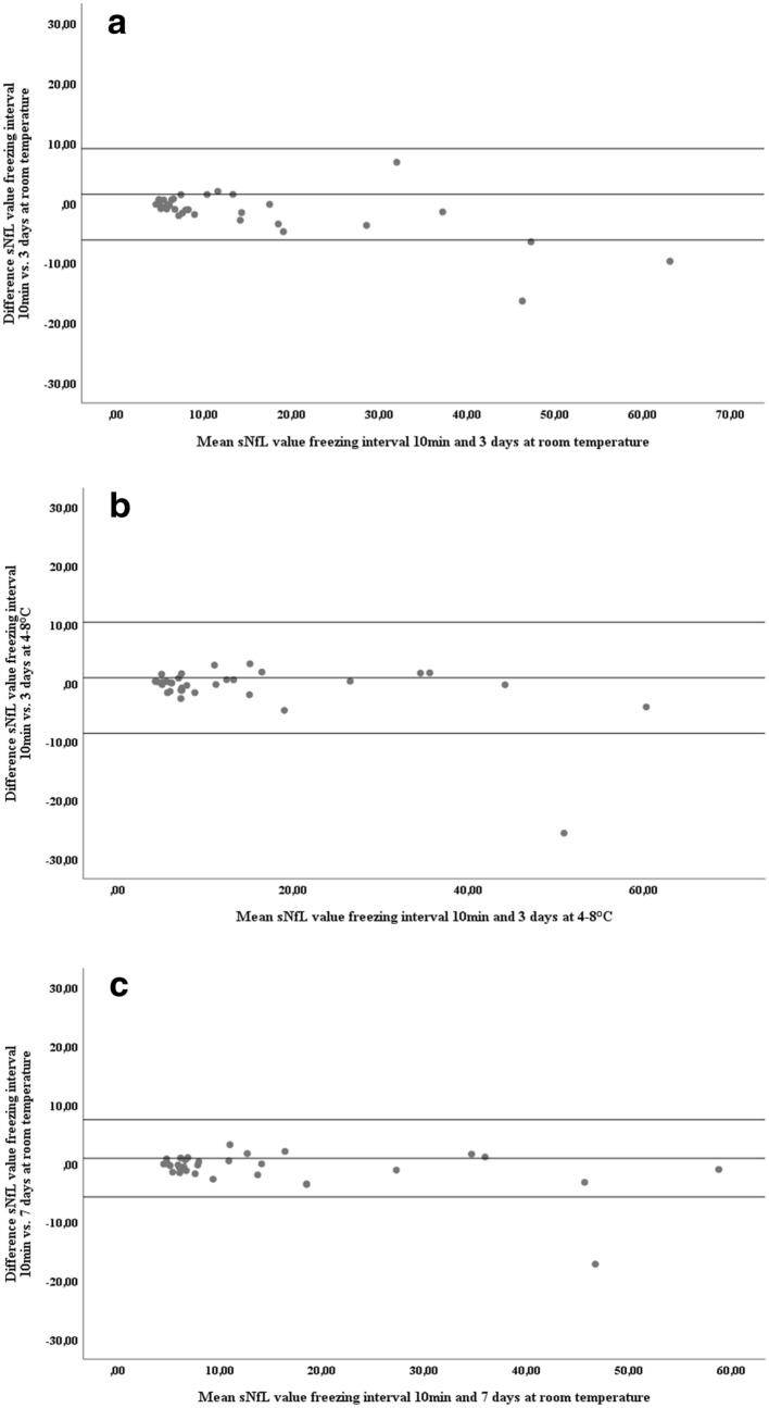
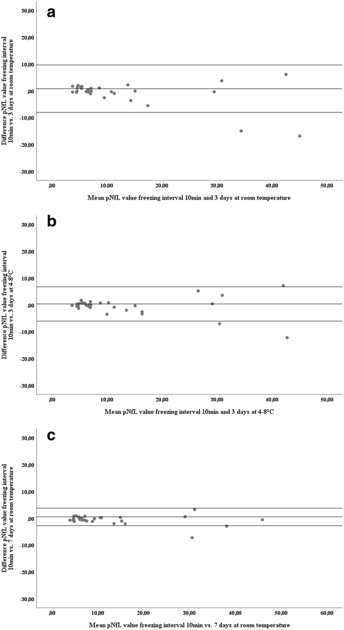
Neurofilament light chain (NfL) has emerged as a biomarker of neuroaxonal damage in several neurologic conditions. With increasing availability of fourth-generation immunoassays detecting NfL in blood, aspects of pre-analytical stability of this biomarker remain unanswered. This study investigated NfL concentrations in serum and plasma samples of 32 patients with neurological diagnoses using state of the art Simoa technology. We tested the effect of delayed freezing of up to 7 days and statistically determined stability and validity of measured concentrations. We found concentrations of NfL in serum and plasma to remain stable at room temperature when processing of samples is delayed up to 7 days (serum: mean absolute difference 0.9 pg/mL, intraindividual variation 1.2%; plasma: mean absolute difference 0.5 pg/mL, intraindividual variation 1.3%). Consistency of these results was nearly perfect for serum and excellent for plasma (intraclass correlation coefficients 0.99 and 0.94, respectively). In conclusion, the soluble serum and plasma NfL concentration remains stable when unprocessed blood samples are stored up to 7 days at room temperature. This information is essential for ensuring reliable study protocols, for example, when shipment of fresh samples is needed.
Conflict of interest statement
There are no direct conflicts of interest with respect to this study. This study was not influenced by a third party. PA has participated in meetings sponsored by, received speaker honoraria or travel funding from Biogen, Merck, Roche, Sanofi-Genzyme and Teva, and received honoraria for consulting from Biogen. He received a research grant from Quanterix International and received funding for the development of a smartphone application for a clinical study from Biogen, Merck, Roche, Sanofi-Genzyme and Teva. MP has nothing to disclose. PSR has received honoraria for consultancy/speaking from AbbVie, Alexion, Almirall, Biogen, Merck, Novartis, Roche, Sandoz, Sanofi-Genzyme, has received research grants from Amicus, Biogen, Merck and Roche. HH received an infrastructural grant from the Austrian Federal Ministry for Science, Research and Education (BBMRI.at) and compensations for Biobanking services in the framework of clinical trials from Glock Health and Blue-Skye Vaccines. PM has nothing to disclose. FL has participated in meetings sponsored by or received honoraria for acting as an advisor/speaker for Bayer, Biogen, Celgene, MedDay, Merck, Novartis, Roche, Sanofi-Genzyme and Teva. AP is a member of the steering committee for the OCTiMS study (Novartis) and ARI network (Zeiss), for which he did not receive any consulting fees. He performed quality control on optical coherence tomography for the Passos study (Novartis), for which he received consulting fees. He received speaker fees from Heidelberg Engineering. CW has nothing to disclose. RL received conference speaker honoraria within the last three years from Bruker BioSpin MR, and support from Siemens Healthcare regarding clinical research using PET/MR. He is a shareholder of the start-up company BM Health GmbH since 2019. TB has participated in meetings sponsored by and received honoraria (lectures, advisory boards, consultations) from pharmaceutical companies marketing treatments for MS: Allergan, Bayer, Biogen, Bionorica, Celgene, MedDay, Merck, Novartis, Octapharma, Roche, Sanofi-Genzyme, Teva. His institution has received financial support in the past 12 months by unrestricted research grants (Biogen, Bayer, Merck, Novartis, Sanofi Aventis, Teva and for participation in clinical trials in multiple sclerosis sponsored by Alexion, Bayer, Biogen, Merck, Novartis, Octapharma, Roche, Sanofi-Genzyme, Teva. HZ has served at scientific advisory boards for Denali, Roche Diagnostics, Wave, Samumed and CogRx, has given lectures in symposia sponsored by Fujirebio, Alzecure and Biogen, and is a co-founder of Brain Biomarker Solutions in Gothenburg AB, a GU Ventures-based platform company at the University of Gothenburg, all unrelated to the work presented in this paper. GB has participated in meetings sponsored by, received speaker honoraria or travel funding from Biogen, Celgene, Merck, Novartis, Sanofi-Genzyme and Teva, and received honoraria for consulting Biogen, Roche and Teva. The other authors report no competing interests or any financial or personal relationships with organizations that could potentially be perceived as influencing the described research.
Figures
References
MeSH terms
Substances
LinkOut - more resources
Full Text Sources
Other Literature Sources
