. 2021 Nov;26(11):6975-6991.
doi: 10.1038/s41380-021-01129-7. Epub 2021 May 27.

Low-intensity ultrasound restores long-term potentiation and memory in senescent mice through pleiotropic mechanisms including NMDAR signaling

Affiliations

Low-intensity ultrasound restores long-term potentiation and memory in senescent mice through pleiotropic mechanisms including NMDAR signaling

Daniel G Blackmore et al. Mol Psychiatry. 2021 Nov.

Abstract

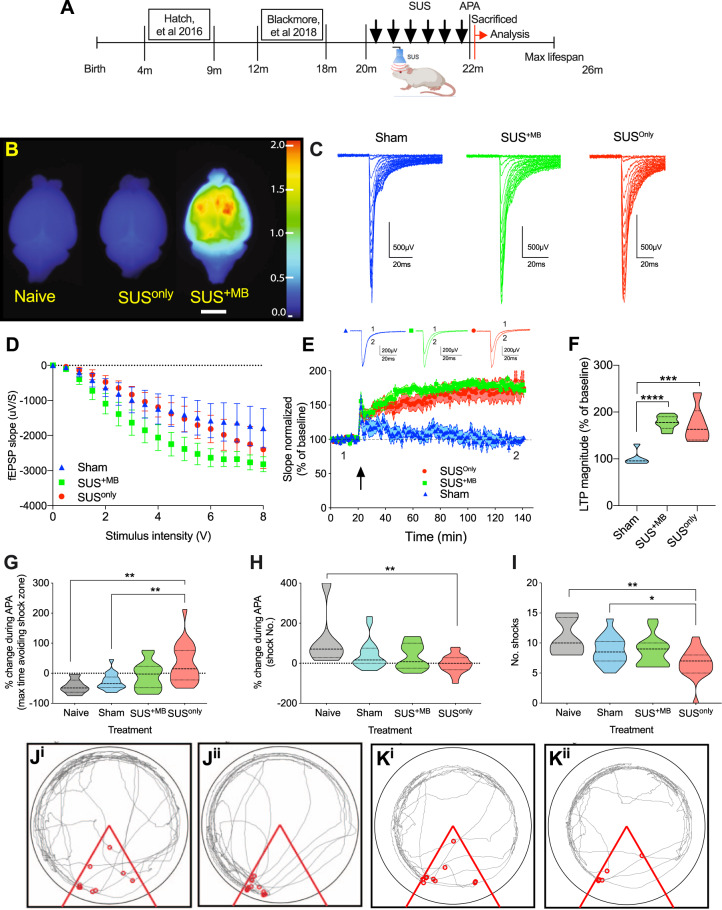
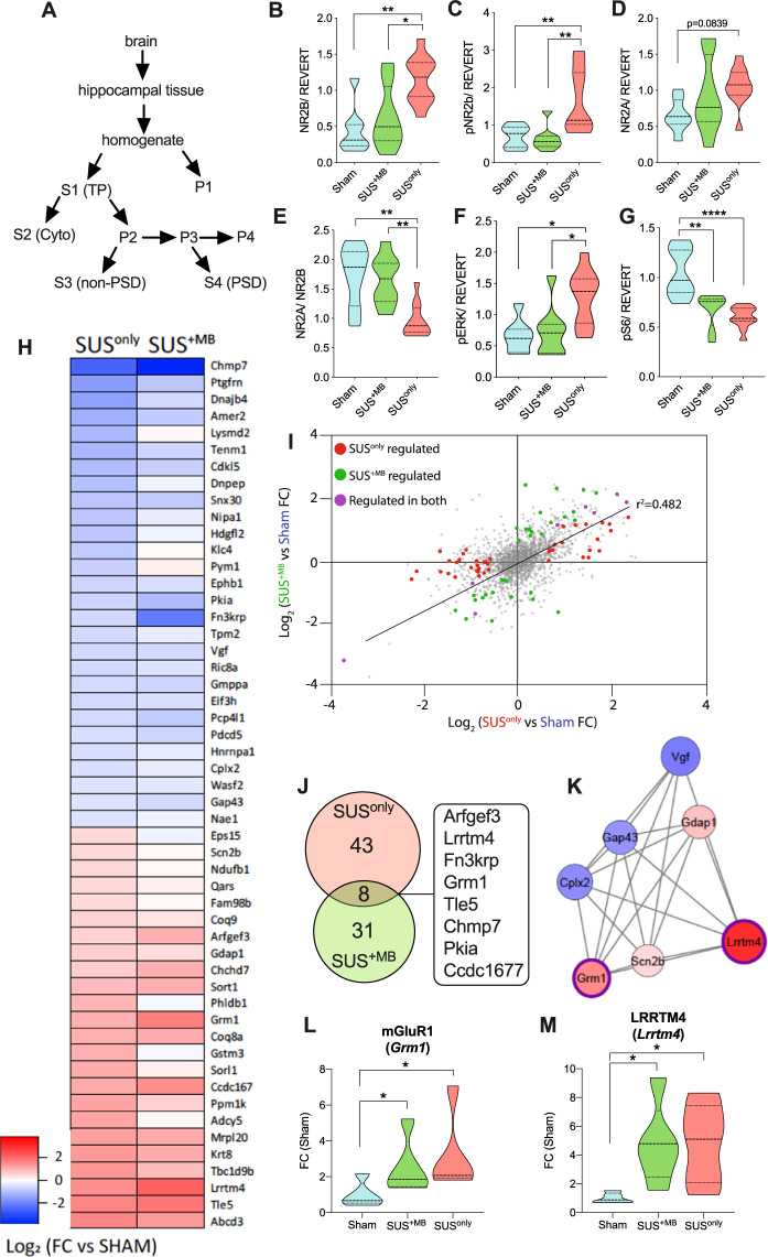
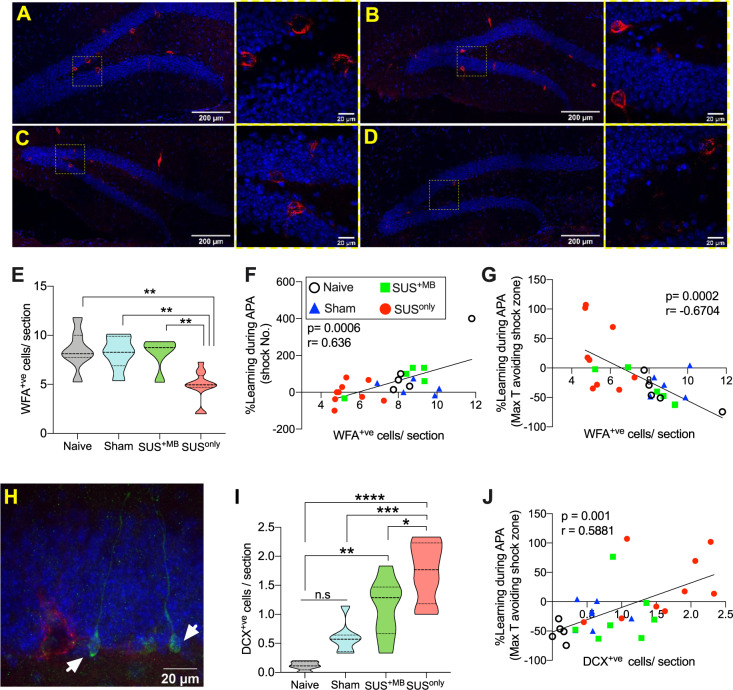
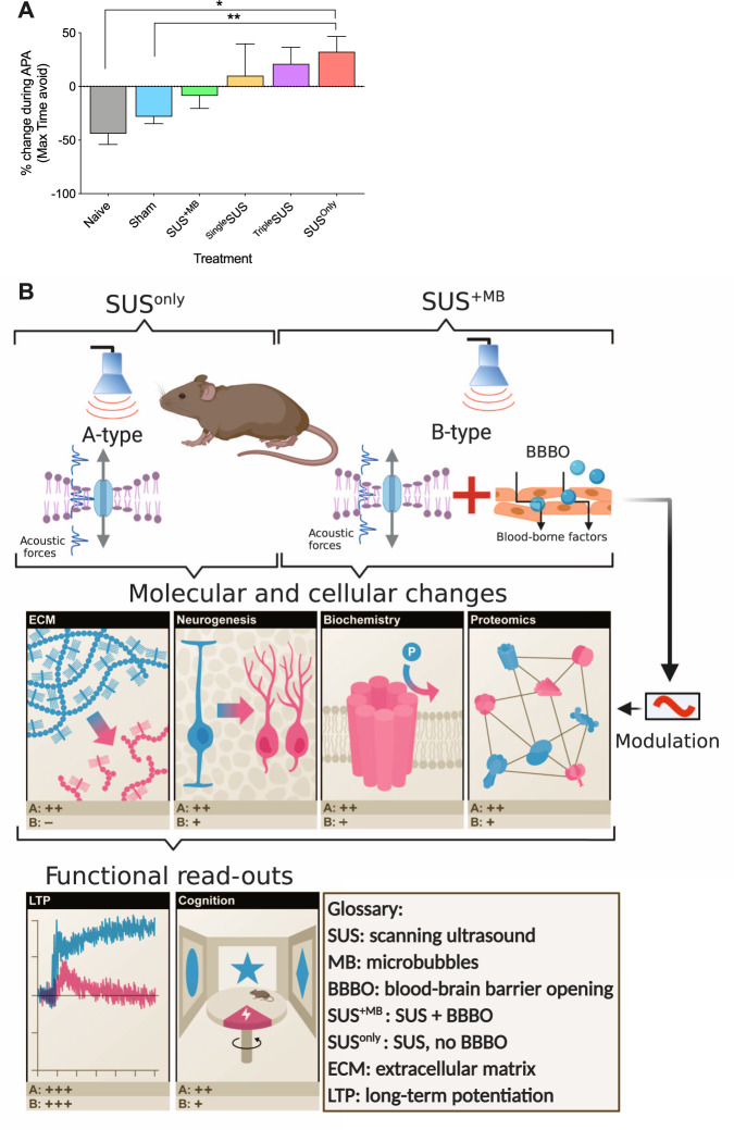
Advanced physiological aging is associated with impaired cognitive performance and the inability to induce long-term potentiation (LTP), an electrophysiological correlate of memory. Here, we demonstrate in the physiologically aged, senescent mouse brain that scanning ultrasound combined with microbubbles (SUS+MB), by transiently opening the blood-brain barrier, fully restores LTP induction in the dentate gyrus of the hippocampus. Intriguingly, SUS treatment without microbubbles (SUSonly), i.e., without the uptake of blood-borne factors, proved even more effective, not only restoring LTP, but also ameliorating the spatial learning deficits of the aged mice. This functional improvement is accompanied by an altered milieu of the aged hippocampus, including a lower density of perineuronal nets, increased neurogenesis, and synaptic signaling, which collectively results in improved spatial learning. We therefore conclude that therapeutic ultrasound is a non-invasive, pleiotropic modality that may enhance cognition in elderly humans.


Conflict of interest statement

The authors declare no competing interests.

Figures

Fig. 1
Fig. 1. The scanning ultrasound equipment accurately delivers treatment and does not generate aversive levels of heat.
A Schematic detail of the sonication procedure, indicating that the transducer sonicates a large area of the mouse brain during each treatment. B Schematic of the movement pattern of the transducer (black arrow). The start and end points are specified, and the transducer was scanned over the area in a raster grid pattern. A typical whole brain sonication used a 5 × 6 spot sonication pattern, covering virtually all of the cerebrum. Each mouse was rotated 180° for subsequent treatments. C To highlight the specificity of the sonication process, less than half of the brain was scanned using a 2 × 6 spot sonication path. The illustration shows a mouse brain following such a SUS+MB sonication to regionally open the BBB, with Evans blue extravasation visualizing this scanning pattern. D The brain from (C) cut coronally in 1 mm sections, with the Evans blue staining showing that ultrasound penetrated through the entirety of the brain. E Representative temperature versus time plot for the sonication using a gray matter mimetic. Note the temperature rise beginning at ~40 s of the elapsed time (red arrow), and the rapid decline after the 6 s sonication ended (black arrow). F Average temperature change (ΔT °C) measured at the skull and with gray matter mimetic 6 s after sonication (n = 6, mean ± SD).
Fig. 2
Fig. 2. SUS treatment rescues LTP in the dentate gyrus of senescent animals and increases synaptic activity, and improves spatial learning ability.
A Schematic highlighting the age of mice previously examined following SUS treatment, with this study examining the effect of SUS on aged, sedentary mice. After the final SUS treatment, animals underwent a single 30 min test on the active place avoidance (APA) test, comparing 22-month-old SUS+MB, SUSonly, sham, and age-matched naive mice. B Naive mice or those that had undergone the SUSonly or SUS+MB procedure were intravenously injected with Evans blue to visualize BBB opening. Whole mouse brains were scanned at 700 nm in the LiCor Odyssey scanner. The heat map shows relative fluorescence intensities, with higher intensities (see color bar) representing BBB opening. Scale bar: 5 mm. C Representative example of an input/output (I/O) curve for sham (mean = −944.9 ± 138.8 µV/s SEM), SUS+MB (mean = −1762 ± 236.7 µV/s SEM), and SUSonly mice (mean = −1164 ± 197.1 µV/s SEM). D SUS treatment increased synaptic transmission only in the SUS+MB group (one-way ANOVA [2.15 = 4.705, p = 0.0137, with Bonferroni post hoc analysis). E Following theta-burst stimulation (TBS) in vitro (indicated by a black arrow), no LTP was observed in the sham mice; however, LTP was fully rescued in both the SUS+MB and SUSonly mice. F Histogram representing the average of the last 10 min of LTP for each treatment represented as a percentage of baseline, with significant increases observed in both the SUS+MB and SUSonly groups (one-way ANOVA [F(2,17) = 19.85, p < 0.001], with Bonferroni post hoc analysis). *p < 0.05, **p < 0.01, ***p < 0.001, and ****p < 0.0001. G The SUSonly group exhibited an increase in the maximum time spent avoiding the aversive shock zone during testing relative to the naive, sham, and SUS+MB treatment groups (one-way ANOVA [F(3,56) = 6.934), p = 0.0005], with Bonferroni post hoc analysis). H The SUSonly group had a significant reduction in the number of shocks in the last 5 min of testing relative to the first 5 min (one-way ANOVA [F(3,56) = 4.552), p = 0.0063], with Bonferroni post hoc analysis). I There was a significant reduction in the number of shocks the SUSonly group received compared to the sham group (one-way ANOVA [F(3,56) = 5.739), p = 0.001], with Bonferroni post hoc analysis). J Representative trace maps of a naive animal for the first 5 min (Ji) and final 5 min (Jii) of the APA test, with red dots representing shocks received. K Representative trace maps of a SUSonly animal during the first 5 min (Ki) and final 5 min (Kii) of the APA test. During the testing period there was a reduction in the number of shocks received following SUSonly treatment.
Fig. 3
Fig. 3. SUS treatment alters the levels and activities of hippocampal proteins in the postsynaptic density fraction and alters the proteomic profile of hippocampal proteins in the total protein fraction.
A Schematic illustrating how specific fractions were obtained from hippocampal samples to conduct biochemical analyses. B NR2B and C pNR2B were increased in postsynaptic fractions from SUSonly mice compared to the sham and SUS+MB groups. D NR2A levels were not altered following SUS treatment. E The NR2A/2B ratio was reduced in SUSonly-treated animals. F There was a significant increase in pERK for SUSonly animals. G Both SUS+MB and SUSonly treatments produced decreases in pS6 levels relative to sham-treated animals (one-way ANOVA with Bonferroni post hoc analysis). H Heat map of proteins significantly altered following SUSonly treatment (|FC| ≥ 1.5, p ≤ 0.05). I Comparison of SUS+MB and SUSonly treatment groups reveals that the vast majority of regulated proteins (72/82) show similar directional changes in fold-change relative to sham controls (Pearson’s correlation, r2 = 0.482). J Venn diagram of proteins significantly altered in abundance in SUS+MB and SUSonly treatment groups compared to sham controls (|FC| ≥ 1.5, p ≤ 0.05). K STRING analysis revealed a cluster associated with synaptic signaling, including increases in both mGluR1 (L) and Lrrtm4 (M), after both SUS+MB and SUSonly treatment (one-way ANOVA with Bonferroni post hoc analysis). *p < 0.05, **p < 0.01, and ****p < 0.0001.
Fig. 4
Fig. 4. SUS treatment reduces the extracellular matrix and increases neurogenesis in aged animals.
AD WFA staining (in red, DAPI in blue) of brain sections from naive (A), sham (B), SUS+MB (C), and SUSonly mice (D). E There was a significant decrease in WFA+ve cells in the dentate gyrus of animals treated with SUSonly (one-way ANOVA [F(3,27) = 9.963, p = 0.0001], with Bonferroni post hoc analysis). F A significant positive correlation was observed between the number of WFA+ve cells and learning ability in the APA task and the change in shock numbers (G). H Representative photomicrograph showing DCX+ve cells (in green, WFA in red) in the dentate gyrus of naive animals. I SUS+MB mice exhibited an increase in the number of DCX+ve cells/section compared to naive animals, whereas SUSonly animals had significantly more DCX+ve cells/section than all other treatment groups (one-way ANOVA [F(3,23) = 18.37, p < 0.0001], with Bonferroni post hoc analysis). J There was a significant positive correlation between the number of DCX+ve cells/section and learning ability in the APA task. *p < 0.05, **p < 0.01, ***p < 0.001, and ****p < 0.0001.
Fig. 5
Fig. 5. Timecourse of memory improvements after SUSonly treatment and integrated model of the effects of SUS on the aging brain.
A Comparing the change in the maximum time animals can avoid the shock zone as a measure of the learning ability during APA reveals a trend toward increased ability after one (SingleSUS, n = 8) and three SUSonly treatments (TripleSUS, n = 8), which after six SUSonly sessions (SUSonly, n = 20) becomes significant (one-way ANOVA [F(5,70) = 3.879, p = 0.0037] with Bonferroni post hoc analysis). B Integrated model differentiating A-type effects of SUS due to a pressure wave (SUSonly) and B-type effects, adding modulatory effects due to BBB opening and the uptake of blood-borne factors by the brain (SUS+MB). The acoustic forces from SUS alter the hippocampal milieu by reducing the ECM, increasing neurogenesis and altering the activity and proteomic profile of hippocampal proteins. These changes ultimately result in improved functional outcomes including reinstatement to induce LTP and improved spatial learning. Scoring (pluses and minuses) indicates the corresponding effect for the respective read-outs.

Similar articles

Cited by

References

    1. Lopez-Otin C, Blasco MA, Partridge L, Serrano M, Kroemer G. The hallmarks of aging. Cell. 2013;153:1194–217. - PMC - PubMed
    1. Blackmore DG, Turpin F, Mohamed AZ, Zong F, Pandit R, Pelekanos M, et al. Multimodal analysis of aged wild-type mice exposed to repeated scanning ultrasound treatments demonstrates long-term safety. Theranostics. 2018;8:6233–47.. - PMC - PubMed
    1. Leinenga G, Götz J. Scanning ultrasound removes amyloid-beta and restores memory in an Alzheimer’s disease mouse model. Sci Tran Med. 2015;7:278ra33. - PubMed
    1. Nisbet RM, Van der Jeugd A, Leinenga G, Evans HT, Janowicz PW, Götz J. Combined effects of scanning ultrasound and a tau-specific single chain antibody in a tau transgenic mouse model. Brain. 2017;140:1220–30.. - PMC - PubMed
    1. Leinenga G, Langton C, Nisbet R, Götz J. Ultrasound treatment of neurological diseases—current and emerging applications. Nat Rev Neurol. 2016;12:161–74. - PubMed

Publication types

Substances