

"Picture-in-Picture" Artifact: Introduction and Characterization of a Hitherto Unrecognized Imaging Artifact in Creating Perfusion Defects in Myocardial Perfusion Single-Photon Emission Computed Tomography
- PMID: 34040303
- PMCID: PMC8130697
- DOI: 10.4103/ijnm.IJNM_55_20
"Picture-in-Picture" Artifact: Introduction and Characterization of a Hitherto Unrecognized Imaging Artifact in Creating Perfusion Defects in Myocardial Perfusion Single-Photon Emission Computed Tomography
Abstract
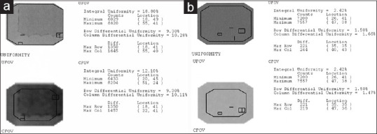
Following a moving hot spot in the projections of raw images and profound perfusion defects in myocardial perfusion single-photon emission computed tomography (SPECT) imaging of a patient, a hypothesis was postulated that the perfusion defects were artifactual, and the high activity concentration of the gallbladder may be a culprit for this phenomenon, owing to flawed event positioning function of the gamma camera due to a malfunctioning digital event processor electronics board. To depict the characteristics of this artifact, a point source containing an activity of 3 mCi of pertechnetate is placed on the scanning table with the detector facing the table (at a distance of 30 cm), and then, in other detector positions and 1-min static images are acquired accordingly. The ratio is calculated as follows: count of the artifactual focus: 1860, count of the index focus: 705,727, and artifactual-to-index focus ratio: 0.003. In testing the uniformity of gamma camera based on the National Electrical Manufacturers Association protocol, a nonuniform response was detected, seemingly, a smaller field of view (FOV) is reproduced in the main FOV causing nonuniformity more than the acceptable level. The smaller flood image lies in the upper right corner of the main flood image. In essence, the extremely bright gallbladder was the source of error, and its image was reproduced in the FOV, which was superimposed on the left ventricular myocardium in some of the projections and was propagated to SPECT images.
Keywords: Gamma camera; imaging artifact; myocardial perfusion single-photon emission computed tomography; perfusion defects; picture-in-picture artifact.
Copyright: © 2021 Indian Journal of Nuclear Medicine.
Conflict of interest statement
There are no conflicts of interest.
Figures


Similar articles
-
The prevalence of a false-positive myocardial perfusion stress SPET test in a skinny patient, induced by projection truncation.Hell J Nucl Med. 2015 Jan-Apr;18(1):79-80. doi: 10.1967/s002449910169. Hell J Nucl Med. 2015. PMID: 25679080
-
Intrinsic uniformity requirements for pinhole SPECT.J Nucl Med Technol. 2006 Mar;34(1):43-7. J Nucl Med Technol. 2006. PMID: 16517968
-
Quantitative assessment of motion artifacts and validation of a new motion-correction program for myocardial perfusion SPECT.J Nucl Med. 2001 May;42(5):687-94. J Nucl Med. 2001. PMID: 11337561
-
Utility of left lateral supine position for myocardial perfusion single-photon emission computed tomography compared with other methods of correcting inferior wall attenuation.Nucl Med Commun. 2015 Mar;36(3):268-78. doi: 10.1097/MNM.0000000000000237. Nucl Med Commun. 2015. PMID: 25356619 Clinical Trial.
-
What is this image? 2019: Image 4 result : Starburst artifact from a remote intense hot spot creating perfusion defects in the LV myocardium on myocardial perfusion SPECT.J Nucl Cardiol. 2020 Feb;27(1):15-17. doi: 10.1007/s12350-019-01998-0. J Nucl Cardiol. 2020. PMID: 31858423 No abstract available.
Cited by
-
"Picture-in-Picture" Artifact in Post-therapeutic 131I Whole-body Survey: Deceiving Spot View but Unraveling Whole-body Scanning.Mol Imaging Radionucl Ther. 2022 Jun 27;31(2):142-144. doi: 10.4274/mirt.galenos.2021.65882. Mol Imaging Radionucl Ther. 2022. PMID: 35771034 Free PMC article.
References
-
- Performance Measurements of Gamma Cameras. Vol. 1. Rosslyn, Virginia: National Electrical Manufacturers Association Standards Publication NU; 2018. [Last accessed on 2020 Dec 19]. National Electrical Manufacturers Association. Available from: https://www.nema.org/Standards/Pages/Performance-Measurements-of-Gamma-C... .
-
- Saha GB, editor. Physics and Radiobiology of Nuclear Medicine. New Yorkd: Springer; 2013. Performance parameters of gamma cameras; pp. 127–51.
-
- Sokole EB, Płachcínska A, Britten A, Georgosopoulou ML, Tindale W, Klett R; EANM Physics Committee, EANM Working Group on Nuclear Medicine Instrumentation Quality Control. Routine quality control recommendations for nuclear medicine instrumentation. Eur J Nucl Med Mol Imaging. 2010;37:662–71. - PubMed
-
- Vienna: International Atomic Energy Agency; 2003. [Last accessed 2020 Feb 01]. International Atomic Energy Agency Quality Control Atlas for Scintillation Camera Systems. Available from: http://www-pub.iaea.org/MTCD/ Publications/PDF/Pub1141_web.pdf .
LinkOut - more resources
Full Text Sources
Other Literature Sources