

MiRNA-29c-3p Promotes Intestinal Inflammation via Targeting Leukemia Inhibitory Factor in Ulcerative Colitis
- PMID: 34040415
- PMCID: PMC8140949
- DOI: 10.2147/JIR.S302832
MiRNA-29c-3p Promotes Intestinal Inflammation via Targeting Leukemia Inhibitory Factor in Ulcerative Colitis
Abstract
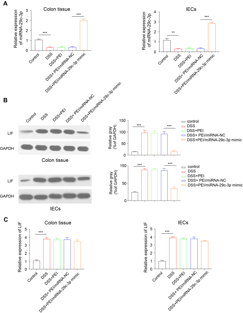
Background: Dysregulation of micro-RNAs (miRNAs) is profoundly linked to inflammatory bowel diseases (IBD), but little is known about the specific biological functions of miRNAs in IBD. This study sought to elucidate the effect and the underlying target of miR-29c-3p in ulcerative colitis (UC).
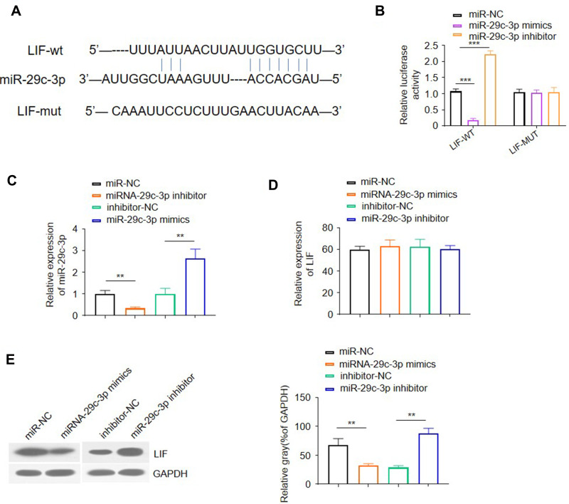
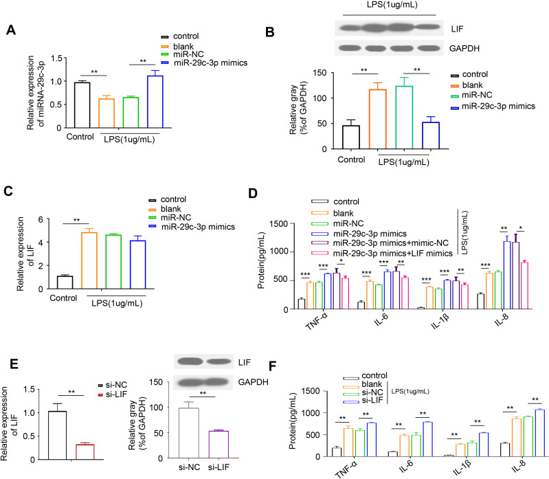
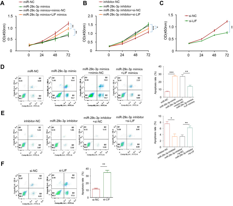
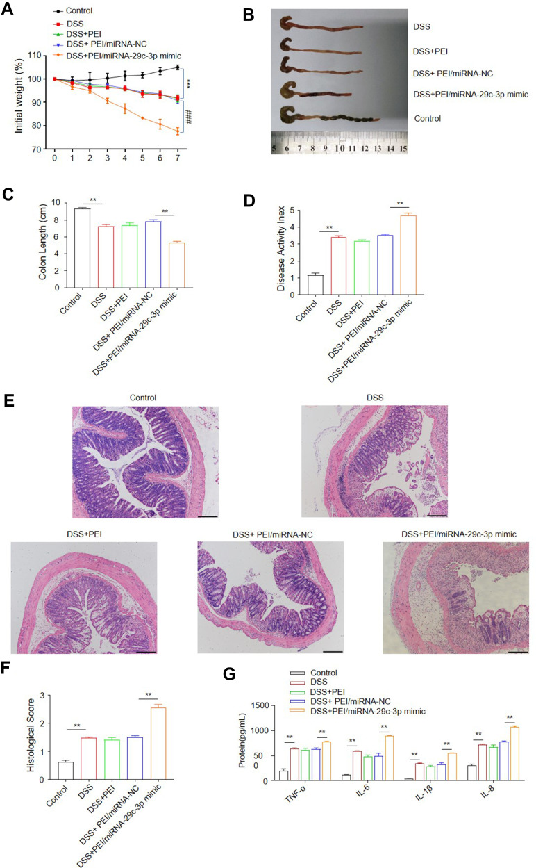
Methods: The levels of miR-29c-3p and leukemia inhibitory factor (LIF) were measured in inflamed lesions of UC patients and dextran sulfate sodium (DSS)-induced colitis mice by quantitative real-time polymerase chain reaction (qRT-PCR) and Western blotting. MiR-29c-3p was predicted to target LIF by bioinformatics software, which was verified via luciferase reporter assay and transfection of miR-29c-3p mimics or inhibitor. The role of miR-29c-3p/LIF axis in intestinal inflammation was explored in experimental colitis mice and Caco-2 cells.
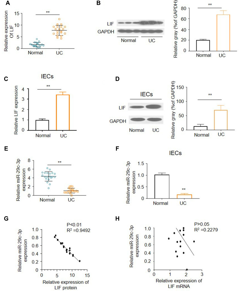
Results: MiR-29c-3p was markedly downregulated while LIF was upregulated in colon tissues of UC patients and DSS-challenged colitis mice as well as in primary intestinal epithelial cells (IECs) and LPS-treated Caco-2 cells. MiR-29c-3p inhibited LIF expression at the transcriptional level via binding to LIF 3'-untranslated region (UTR) in Caco-2 cells. Targeting miR-29c-3p/LIF axis regulated inflammatory cytokines production, cell proliferation and apoptosis. Overexpression of miR-29c-3p aggravated mice experimental colitis via suppressing LIF.
Conclusion: Our findings demonstrate that the upregulation of miR-29c-3p promotes gut inflammation and the expression of pro-inflammatory mediators via suppressing LIF, thereby modulating the pathogenesis of UC.
Keywords: apoptosis; inflammation; leukemia inhibitory factor; microRNA-29c-3p; ulcerative colitis.
© 2021 Guo et al.
Conflict of interest statement
The authors declare that there are no conflicts of interest.
Figures
Similar articles
-
miR-29c-3p Accelerates Mucosal Repair in Dextran Sodium Sulfateinduced Ulcerative Colitis Mice through the KDM6B/H3K27me3/LDHA Axis.Protein Pept Lett. 2023;30(6):459-468. doi: 10.2174/0929866530666230511115213. Protein Pept Lett. 2023. PMID: 37171009
-
Long Noncoding RNA X-Inactive-Specific Transcript Promotes the Secretion of Inflammatory Cytokines in LPS Stimulated Astrocyte Cell Via Sponging miR-29c-3p and Regulating Nuclear Factor of Activated T cell 5 Expression.Front Endocrinol (Lausanne). 2021 Mar 12;12:573143. doi: 10.3389/fendo.2021.573143. eCollection 2021. Front Endocrinol (Lausanne). 2021. PMID: 33776905 Free PMC article.
-
miR-141-3p alleviates ulcerative colitis by targeting SUGT1 to inhibit colonic epithelial cell pyroptosis.Autoimmunity. 2023 Dec;56(1):2220988. doi: 10.1080/08916934.2023.2220988. Autoimmunity. 2023. PMID: 37317573
-
Suppression of microRNA-222-3p ameliorates ulcerative colitis and colitis-associated colorectal cancer to protect against oxidative stress via targeting BRG1 to activate Nrf2/HO-1 signaling pathway.Front Immunol. 2023 Jan 27;14:1089809. doi: 10.3389/fimmu.2023.1089809. eCollection 2023. Front Immunol. 2023. PMID: 36776858 Free PMC article.
-
Suppression of miR-330-3p alleviates DSS-induced ulcerative colitis and apoptosis by upregulating the endoplasmic reticulum stress components XBP1.Hereditas. 2020 May 9;157(1):18. doi: 10.1186/s41065-020-00135-z. Hereditas. 2020. PMID: 32386518 Free PMC article.
Cited by
-
RNA-Seq Reveals the Role of miR-29c in Regulating Inflammation and Oxidative Stress of Bovine Mammary Epithelial Cells.Front Vet Sci. 2022 Apr 1;9:865415. doi: 10.3389/fvets.2022.865415. eCollection 2022. Front Vet Sci. 2022. PMID: 35433915 Free PMC article.
-
Regulatory Role of IGF2BP2 in Intestinal Mucosal Barrier Dysfunction in Ulcerative Colitis.Turk J Gastroenterol. 2025 Jan 6;36(4):269-279. doi: 10.5152/tjg.2025.24192. Turk J Gastroenterol. 2025. PMID: 40241612 Free PMC article.
-
A collection of patient-derived intestinal organoid lines reveals epithelial phenotypes associated with genetic drivers of pediatric inflammatory bowel disease.bioRxiv [Preprint]. 2025 Jun 16:2025.06.11.659052. doi: 10.1101/2025.06.11.659052. bioRxiv. 2025. PMID: 40666956 Free PMC article. Preprint.
-
Modulation of the Inflammatory Response in Polycystic Ovary Syndrome (PCOS)-Searching for Epigenetic Factors.Int J Mol Sci. 2022 Nov 24;23(23):14663. doi: 10.3390/ijms232314663. Int J Mol Sci. 2022. PMID: 36498989 Free PMC article. Review.
-
An Integrative Approach to the Current Treatment of HIV-Associated Neurocognitive Disorders and the Implementation of Leukemia Inhibitor Factor as a Mediator of Neurocognitive Preservation.Life (Basel). 2023 Nov 11;13(11):2194. doi: 10.3390/life13112194. Life (Basel). 2023. PMID: 38004334 Free PMC article. Review.
References
LinkOut - more resources
Full Text Sources
Other Literature Sources
