. 2021 May 10:13:673210.
doi: 10.3389/fnsyn.2021.673210. eCollection 2021.

Impact of Perineuronal Nets on Electrophysiology of Parvalbumin Interneurons, Principal Neurons, and Brain Oscillations: A Review

Affiliations

Impact of Perineuronal Nets on Electrophysiology of Parvalbumin Interneurons, Principal Neurons, and Brain Oscillations: A Review

Jereme C Wingert et al. Front Synaptic Neurosci. .

Abstract

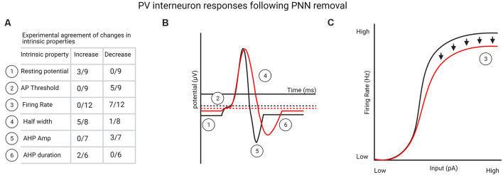
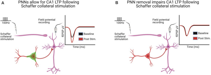
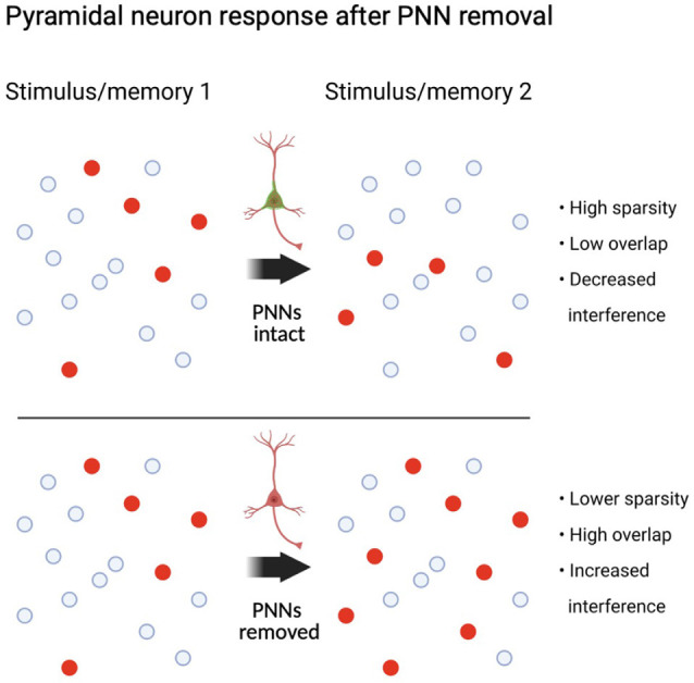
Perineuronal nets (PNNs) are specialized extracellular matrix structures that surround specific neurons in the brain and spinal cord, appear during critical periods of development, and restrict plasticity during adulthood. Removal of PNNs can reinstate juvenile-like plasticity or, in cases of PNN removal during early developmental stages, PNN removal extends the critical plasticity period. PNNs surround mainly parvalbumin (PV)-containing, fast-spiking GABAergic interneurons in several brain regions. These inhibitory interneurons profoundly inhibit the network of surrounding neurons via their elaborate contacts with local pyramidal neurons, and they are key contributors to gamma oscillations generated across several brain regions. Among other functions, these gamma oscillations regulate plasticity associated with learning, decision making, attention, cognitive flexibility, and working memory. The detailed mechanisms by which PNN removal increases plasticity are only beginning to be understood. Here, we review the impact of PNN removal on several electrophysiological features of their underlying PV interneurons and nearby pyramidal neurons, including changes in intrinsic and synaptic membrane properties, brain oscillations, and how these changes may alter the integration of memory-related information. Additionally, we review how PNN removal affects plasticity-associated phenomena such as long-term potentiation (LTP), long-term depression (LTD), and paired-pulse ratio (PPR). The results are discussed in the context of the role of PV interneurons in circuit function and how PNN removal alters this function.

Keywords: memory; oscillations; parvalbumin; perineuronal nets (PNNs); plasticity.


Conflict of interest statement

The authors declare that the research was conducted in the absence of any commercial or financial relationships that could be construed as a potential conflict of interest.

Figures

Figure 1
Figure 1
(A) List of perineuronal net (PNN) removal-mediated changes in parvalbumin (PV) interneuron intrinsic properties that one-third or more of the experiments agreed upon (see Supplementary Table 1 for a complete list of studies). (B) Graphical representation from table shown in (A) of changes in PV interneuron intrinsic properties following PNN removal. (C) Experimentally agreed upon firing rate changes to PV interneurons following PNN removal, indicating deficits in firing rate at high stimulation levels. Created with BioRender.com.
Figure 2
Figure 2
Hippocampal long-term potentiation (LTP) in the CA1 following Schaffer collateral stimulation was altered in six of the seven studies following PNN removal [see Supplementary Table 3 for a complete list of LTP/long-term depression (LTD) and paired-pulse ratio (PPR) studies]. (A) With PNNs intact, Schaffer collateral stimulation induces LTP. (B) With PNNs removed, Schaffer collateral stimulation-induced LTP is impaired. Created with BioRender.com.
Figure 3
Figure 3
Circuit state following PV interneuron dysfunction after PNN removal. Neural assemblies originally representing a stimulus or memory may overlap when exposed to a similar stimulus or memory if PNNs are removed between the first and second exposure. Summary is based on PV interneuron specific manipulations and circuit functions (see “Role of PV Interneurons in Pattern Separation and Sparse Encoding” section). Created with BioRender.com.

Similar articles

Cited by

References

    1. Abbas A. I., Sundiang M. J. M., Henoch B., Morton M. P., Bolkan S. S., Park A. J., et al. . (2018). Somatostatin interneurons facilitate hippocampal-prefrontal synchrony and prefrontal spatial encoding. Neuron 100, 926.e3–939.e3. 10.1016/j.neuron.2018.09.029 - DOI - PMC - PubMed
    1. Agetsuma M., Hamm J. P., Tao K., Fujisawa S., Yuste R. (2018). Parvalbumin-positive interneurons regulate neuronal ensembles in visual cortex. Cereb. Cortex 28, 1831–1845. 10.1093/cercor/bhx169 - DOI - PMC - PubMed
    1. Allen W. E., Kauvar I. V., Chen M. Z., Richman E. B., Yang S. J., Chan K., et al. . (2017). Global representations of goal-directed behavior in distinct cell types of mouse neocortex. Neuron 94, 891.e6–907.e6. 10.1016/j.neuron.2017.04.017 - DOI - PMC - PubMed
    1. Atallah B. V., Bruns W., Carandini M., Scanziani M. (2012). Parvalbumin-expressing interneurons linearly transform cortical responses to visual stimuli. Neuron 73, 159–170. 10.1016/j.neuron.2011.12.013 - DOI - PMC - PubMed
    1. Avermann M., Tomm C., Mateo C., Gerstner W., Petersen C. C. (2012). Microcircuits of excitatory and inhibitory neurons in layer 2/3 of mouse barrel cortex. J. Neurophysiol. 107, 3116–3134. 10.1152/jn.00917.2011 - DOI - PubMed

LinkOut - more resources