

Eccentric saccular aneurysm formation of the infrarenal aorta from an arterial wall tear
- PMID: 34040688
- PMCID: PMC8144532
- DOI: 10.1016/j.radcr.2021.04.046
Eccentric saccular aneurysm formation of the infrarenal aorta from an arterial wall tear
Erratum in
-
Erratum to "Eccentric saccular aneurysm formation of the infrarenal aorta from an arterial wall tear" [Radiology Case Reports 16 (2021) 1854-1856].Radiol Case Rep. 2022 Apr 12;17(6):2287. doi: 10.1016/j.radcr.2022.03.093. eCollection 2022 Jun. Radiol Case Rep. 2022. PMID: 35574568 Free PMC article.
-
Erratum regarding missing patient consent statements in previously published articles.Radiol Case Rep. 2023 Jan 20;18(3):1383-1384. doi: 10.1016/j.radcr.2022.10.047. eCollection 2023 Mar. Radiol Case Rep. 2023. PMID: 36818994 Free PMC article.
Abstract
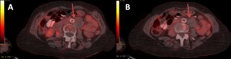
Eccentric saccular aneurysms result from a focal weakness of the arterial wall that may be due to a focal tear or a partial disruption of the arterial wall. Saccular morphology itself is often used as a factor for immediate intervention, because the risk of rupture is higher than that of the common fusiform aneurysms. We present a case of a 72-year-old female patient with a huge saccular aneurysm of the infrarenal aorta. In this case report, we discuss the algorithm that can be used for the differential diagnosis of any saccular shape aneurysm and that the main parameter that needs to be clarified before the endovascular treatment of any saccular aneurysm is the presence or absence of infection of the arterial wall.
Keywords: Endovascular treatment; Infected aneurysm; Saccular aneurysm.
© 2021 The Authors. Published by Elsevier Inc. on behalf of University of Washington.
Figures


References
-
- Eggebrecht H., Plicht B., Kahlert P., Erbel R. Intramural hematoma and penetrating ulcers: indications to endovascular treatment. Eur J Vasc Endovasc Surg. 2009;38:659–665. - PubMed
-
- Lijnen H.R. Metalloproteinases in development and progression of vascular disease. Pathophysiol Haemost Thromb. 2003;33:275–281. - PubMed
-
- Brown S.L., Busuttil R., Baker J., Machleder H., Moore W., Barker W. Bacteriologic and surgical determinants of survival in patients with mycotic aneurysms. J Vasc Surg. 1984;1:541–547. - PubMed
-
- Spacek M., Stadler P., Bĕlohlávek O., Sebesta P. Contribution to FDG-PET/CT diagnostics and post-operative monitoring of patients with mycotic aneurysm of the thoracic aorta. Acta Chir Belg. 2010;110(1):106–108. - PubMed
-
- Walsh D.W., Ho V., Haggerty M. Mycotic aneurysm of the aorta: MRI and MRA features. J Magn Reson Imag. 1997;7(2):312–315. - PubMed
Publication types
LinkOut - more resources
Full Text Sources
Other Literature Sources
